Page 302 - Feline Cardiology
P. 302
310  Section H: Arterial Thromboembolism


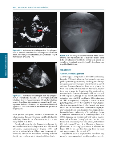
                                     AO


                                    LA


              Figure 20.5.  A	short-axis	echocardiogram	from	the	right	para-
              sternum	obtained	at	the	heart	base	showing	the	aorta	and	left
              atrium.	Note	the	hypoechoic	thrombus	(arrow)	within	the	body	of	  Figure 20.7.  An	aortogram	obtained	from	a	cat	with	a	“saddle”
        Arterial Thromboembolism                                 to	the	pelvis	because	of	a	clot	in	the	terminal	aorta	(arrow),	and
              the	left	atrium	(LA);	aorta	=	Ao.
                                                                 embolus.	Note	the	contrast	in	the	aortic	blood	is	blocked	cranial
                                                                 no	collateral	circulation	is	present	to	the	pelvic	limbs.	Image	cour-
                                                                 tesy	of	Dr.	Mark	Kittleson.


                                                                 TREATMENT
                                                                 Acute Case Management
                                                                 Acute therapy of ATE patients is directed toward manag-
                                                                 ing pain, CHF, or significant arrhythmias when present;
                                                                 general patient support, notably involving pain manage-
                                                                 ment; and adjunctive therapies to limit thrombus growth
                                                                 or future thrombus formation. In the acute phase, a 24-
                                                                 hour care facility is best suited for these cases, because
                                                                 there may be acute life-threatening deterioration at any
                                                                 time during the first several days after ATE has occurred.
              Figure 20.6.  A	short-axis	echocardiogram	from	the	right	para-  If CHF is present, therapy should be initiated similarly
              sternum	obtained	at	the	heart	base	and	showing	the	aorta	and	  as in other cats with CHF (see Chapter 19 for discussion
              left	atrium.	Note	the	hypoechoic	or	grey	debris	in	the	left	atrium	  of  CHF  management).  Analgesia  is  crucial  in  these
              (arrows).	In	real	time,	this	spontaneous	contrast	is	visible	swirl-  patients, particularly for the first 24 to 48 hours, because
              ing	around	the	left	atrial	chamber	and	represents	red	blood	cell	  after that time period, there is often lack of pain noted
              aggregation.	 Left	 atrial	 smoke	 in	 the	 modified	 left	 parasternal	  in cats with a saddle embolus. In humans with periph-
              long-axis	view.
                                                                 eral vascular disease, axonal degeneration begins after 24
                                                                 hours of ischemia and severity of the pathologic change
              these  patients  (neoplasia,  systemic  inflammation  or   is dependent on the duration of ischemia (Nakuda et al.
              other systemic diseases). Neoplasia was identified as the   1996). Analgesia can be addressed with various medica-
              underlying disease in 5% of the cats with ATE in one   tions such as fentanyl (2–5 µg/kg/hr as a CRI for 12–18
              study (Smith et al. 2003).                         hours until a fentanyl patch takes effect), butorphanol
                 Occasionally, more invasive diagnostic testing may be   (0.1 to 0.2 mg/kg IV every 4 to 6 hours), or buprenor-
              warranted to confirm the diagnosis of ATE. Abdominal   phine (0.005 to 0.015 mg/kg IV every 6 to 8 hours). See
              ultrasound,  angiocardiography  (Figure  20.7),  and   Figure 20.8 for an algorithm breaking down the acute
              nuclear scintigraphy have all been used to evaluate the   and long-term care of a cat with ATE.
              obstructed  site  further.  Procedures  requiring  sedation   Vasodilators,  such  as  acepromazine,  have  been  sug-
              should only be attempted in clinically stable patients.  gested to encourage arterial vasodilation; however, arte-

