Page 300 - Feline Cardiology
P. 300
308  Section H: Arterial Thromboembolism


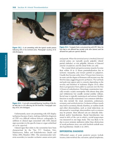
              Figure 20.2.  A	cat	presenting	with	the	typical	caudal	paresis	  Figure 20.4.  Footpads	from	a	cat	presenting	with	ATE.	Note	the
              following	ATE	of	the	terminal	aorta.	Photograph	courtesy	of	Dr.	  foot	that	is	not	affected	has	normal,	pink	color	(above)	and	the
              John	Bonagura.                                     affected	foot	pads	are	cyanotic	(below).
        Arterial Thromboembolism                                 and paresis. When the terminal aorta is involved, femoral


                                                                 arterial  pulses  are  typically  poorly  palpable  (distal/
                                                                 partial  embolus)  or  not  palpable  (absence  of  femoral
                                                                 arterial circulation) and the distal limbs are cold.
                                                                   The cranial tibial and gastrocnemius muscles become
                                                                 firm  within  10  to  12  hours  postembolization  due  to
                                                                 ischemic  myopathy,  and  remain  painful  on  palpation.
                                                                 Usually they become softer 24 to 72 hours later; however,
                                                                 in some cats the degree of firmness will increase over the
                                                                 first few days, suggesting poorer perfusion. The nail beds
                                                                 and pads may appear pale to cyanotic depending on the
                                                                 severity and duration of ischemia (Figure 20.4); often
                                                                 there is progression from pallor to cyanosis over the first
                                                                 12 hours of embolization. Neurologic examination typi-
                                                                 cally reveals a sciatic neuropathy with absent deep pain
                                                                 and  withdrawal,  but  usually  normal  patellar  reflexes.
                                                                 Rectal tone is typically maintained, and urinary bladder
                                                                 function is usually maintained. Less common emboliza-
                                                                 tion  sites  include  the  renal,  mesenteric,  pulmonary,
              Figure 20.3.  A	cat	with	nonweightbearing,	knuckling	of	the	dis-  coronary, and cerebral arteries. Occlusion of these vessels
              tal	limb	due	to	ATE	affecting	the	left	forelimb.	Photograph	cour-  may lead to azotemia, bloody diarrhea, or sudden death
              tesy	of	Dr.	John	Bonagura.                         and  other  abnormalities,  such  as  renal  infarctions  on
                                                                 ultrasound or neurologic abnormalities such as seizures
                                                                 may be present. Most affected cats are clinically dehy-
                 Unfortunately, most cats presenting with ATE display
              tachypnea because of pain, making a definitive diagnosis   drated  and/or  hypothermic.  Rectal  hypothermia  was
              of CHF very difficult without thoracic radiographs. In   noted in 66% of the cats in which a rectal temperature
                                                                 was measured in one large study. This was likely to be
              addition to clinical signs associated with CHF, clinical
              signs secondary to the specific tissue or organ embolized   due to poor systemic perfusion rather than obstruction
                                                                 of rectal perfusion since cats with forelimb emboli were
              are to be expected.
                 Clinical signs that relate to the extremities have been   also hypothermic (Smith et al. 2003).
              characterized  by  the  “five  P’s”:  Paralysis,  Pain,
              Pulselessness,  Pallor,  and  Poikilothermia  (Smith  and   DIFFERENTIAL DIAGNOSIS
              Tobias 2004, Flanders 1986). The neuromuscular isch-  Differential  causes  of  acute  posterior  paresis  include
              emia secondary to vascular occlusion causes severe pain   trauma, intervertebral disc extrusion, spinal lymphoma,

