Page 318 - Feline Cardiology
P. 318
Chapter 21: Systemic Hypertension 327
A A
B B Systemic Hypertension
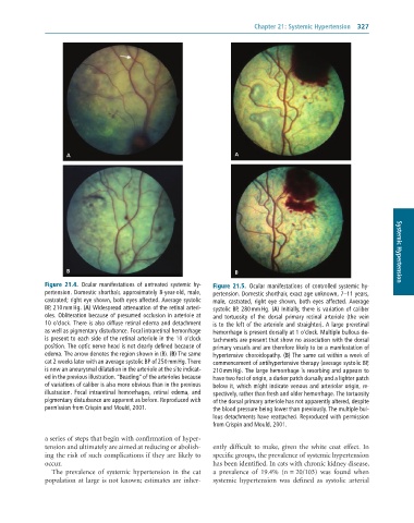
Figure 21.4. Ocular manifestations of untreated systemic hy- Figure 21.5. Ocular manifestations of controlled systemic hy-
pertension. Domestic shorthair, approximately 8-year-old, male, pertension. Domestic shorthair, exact age unknown, 7–11 years,
castrated; right eye shown, both eyes affected. Average systolic male, castrated, right eye shown, both eyes affected. Average
BP, 210 mm Hg. (A) Widespread attenuation of the retinal arteri- systolic BP, 280 mm Hg. (A) Initially, there is variation of caliber
oles. Obliteration because of presumed occlusion in arteriole at and tortuosity of the dorsal primary retinal arteriole (the vein
10 o’clock. There is also diffuse retinal edema and detachment is to the left of the arteriole and straighter). A large preretinal
as well as pigmentary disturbance. Focal intraretinal hemorrhage hemorrhage is present dorsally at 1 o’clock. Multiple bullous de-
is present to each side of the retinal arteriole in the 10 o’clock tachments are present that show no association with the dorsal
position. The optic nerve head is not clearly defined because of primary vessels and are therefore likely to be a manifestation of
edema. The arrow denotes the region shown in (B). (B) The same hypertensive choroidopathy. (B) The same cat within a week of
cat 2 weeks later with an average systolic BP of 250 mm Hg. There commencement of antihypertensive therapy (average systolic BP,
is now an aneurysmal dilatation in the arteriole at the site indicat- 210 mm Hg). The large hemorrhage is resorbing and appears to
ed in the previous illustration. “Beading” of the arterioles because have two foci of origin, a darker patch dorsally and a lighter patch
of variations of caliber is also more obvious than in the previous below it, which might indicate venous and arteriolar origin, re-
illustration. Focal intraretinal hemorrhages, retinal edema, and spectively, rather than fresh and older hemorrhage. The tortuosity
pigmentary disturbance are apparent as before. Reproduced with of the dorsal primary arteriole has not apparently altered, despite
permission from Crispin and Mould, 2001. the blood pressure being lower than previously. The multiple bul-
lous detachments have reattached. Reproduced with permission
from Crispin and Mould, 2001.
a series of steps that begin with confirmation of hyper
tension and ultimately are aimed at reducing or abolish ently difficult to make, given the white coat effect. In
ing the risk of such complications if they are likely to specific groups, the prevalence of systemic hypertension
occur. has been identified. In cats with chronic kidney disease,
The prevalence of systemic hypertension in the cat a prevalence of 19.4% (n = 20/103) was found when
population at large is not known; estimates are inher systemic hypertension was defined as systolic arterial

