Page 319 - Feline Cardiology
P. 319
328 Section I: Systemic Hypertension
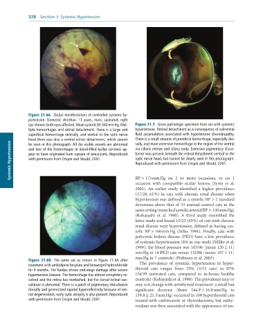
Figure 21.6A. Ocular manifestations of controlled systemic hy-
pertension. Domestic shorthair, 13 years, male, castrated, right
eye shown, both eyes affected. Mean systolic BP, 360 mm Hg. Mul- Figure 21.7. Gross pathologic specimen from cat with systemic
tiple hemorrhages and retinal detachment. There is a large and hypertension. Retinal detachment as a consequence of subretinal
superficial hemorrhage ventrally, and ventral to the optic nerve fluid accumulation associated with hypertensive choroidopathy.
head there was also a ventral retinal detachment, which cannot There is a small amount of preretinal hemorrhage, especially dor-
Systemic Hypertension pear to have originated from rupture of aneurysms. Reproduced bance was present beneath the retinal detachment ventral to the
sally, and more extensive hemorrhage in the region of the ventral
be seen in this photograph. All the visible vessels are abnormal
ora ciliaris retinae and ciliary body. Extensive pigmentary distur-
and two of the hemorrhages in blood-filled bullae (arrows) ap-
optic nerve head, but cannot be clearly seen in this photograph.
with permission from Crispin and Mould, 2001.
Reproduced with permission from Crispin and Mould, 2001.
BP > 175 mm Hg on 2 or more occasions, or on 1
occasion with compatible ocular lesions (Syme et al.
2002). An earlier study identified a higher prevalence
(17/28, 61%) in cats with chronic renal disease when
hypertension was defined as a systolic BP > 2 standard
deviations above that of 33 normal control cats in the
same setting (none had systolic arterial BP > 140 mm Hg)
(Kobayashi et al. 1990). A third study resembled the
latter study and found 15/23 (65%) of cats with chronic
renal disease were hypertensive, defined as having sys
tolic BP > 160 mm Hg (Stiles 1994). Finally, cats with
polycystic kidney disease (PKD) have a low prevalence
of systemic hypertension (0/6 in one study [Miller et al.
1999]; the blood pressure was 145/96 [mean 120 ± 11]
mm Hg in 14 PKD cats versus 132/86 [mean: 107 ± 11]
mm Hg in 7 controls) (Pedersen et al. 2003).
Figure 21.6B. The same cat as shown in Figure 21.6A after
treatment with amlodipine besylate and benazepril hydrochloride The prevalence of systemic hypertension in hyper
for 4 months. The fundus shows end-stage damage after severe thyroid cats ranges from 23% (3/13 cats) to 87%
hypertensive disease. The hemorrhage has almost completely re- (34/39 untreated cats, compared to inhouse healthy
solved and the retina has reattached, but the dorsal retinal vas- controls) (Kobayashi et al. 1990). The prevalence may or
culature is abnormal. There is a patch of pigmentary disturbance may not change with antithyroid treatment: a small but
dorsally and generalized tapetal hyperreflectivity because of reti- significant decrease (from 144.9 ± 16.8 mm Hg to
nal degeneration; early optic atrophy is also present. Reproduced 139.8 ± 21.3 mm Hg) occurred in 100 hyperthyroid cats
with permission from Crispin and Mould, 2001. treated with carbimazole or thyroidectomy, but euthy
roidism was then associated with the appearance of sys

