Page 317 - Feline Cardiology
P. 317
Measure BP
                                                 Identify TOD &
                                                Concurrent diseases

                   BP < 150/95 mmHg             BP ≥ 150/95 mmHg

                   Minimal risk of TOD  Ocular/CNS TOD present  No ocular/CNS TOD present
                 Re-evaluate in 3-6 months


                                         Candidate for     Re-measure BP
                                     initiation or escalation of  within 7 days
                                     antihypertensive therapy

                   BP < 150/95 mmHg  BP 150-159/95-99 mmHg                 BP 160-179/100-119 mmHg  BP ≥ 180/120 mmHg

                     Re-measure BP     Evidence of TOD?                       Evidence of TOD        Yes:
                     in 1-3 months                                              or cause of        Candidate for
                                                                            secondary hypertension?  initiation or escalation of
                                                                                               anti-hypertensive therapy

                                  Yes:               No:                 Yes:                No:
                               Candidate for     Manage causes of     Candidate for  Clnical judgement: Candidate for
                            initation or escalation  secondary hypertension.  initiation or escalation of  anti-hypertensive therapy -or-
                          of anti-hypertensive therapy  Re-measure BP in 1-3 months  anti-hypertensive therapy  re-measure BP in 1 month


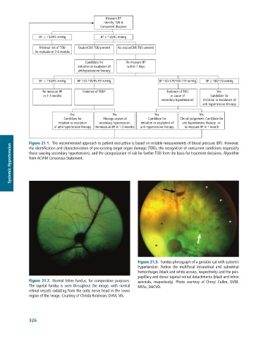
              Figure 21.1.  The	recommended	approach	to	patient	evaluation	is	based	on	reliable	measurements	of	blood	pressure	(BP).	However,
      Systemic Hypertension  those	causing	secondary	hypertension),	and	the	categorization	of	risk	for	further	TOD	form	the	basis	for	treatment	decisions.	Algorithm
              the	identification	and	characterization	of	pre-existing	target	organ	damage	(TOD),	the	recognition	of	concurrent	conditions	(especially
              from	ACVIM	Consensus	Statement.


                                                                                                *

                                                                                                       *


                                                                                                           *
                                                                           *


                                                                         *


                                                                 Figure 21.3.  Fundus	photograph	of	a	geriatric	cat	with	systemic
                                                                 hypertension.	 Notice	 the	 multifocal	 intraretinal	 and	 subretinal
                                                                 hemorrhages	(black	and	white	arrows,	respectively)	and	the	peri-
                                                                 papillary	and	dorsal	tapetal	retinal	detachments	(black	and	white
              Figure 21.2.  Normal	feline	fundus,	for	comparative	purposes.	  asterisks,	 respectively).	 Photo	 courtesy	 of	 Cheryl	 Cullen,	 DVM,
              The	tapetal	fundus	is	seen	throughout	the	image,	with	normal	  MVSc,	DACVO.
              retinal	vessels	radiating	from	the	optic	nerve	head	in	the	lower
              region	of	the	image.	Courtesy	of	Christa	Robinson,	DVM,	MS.


              326

