Page 344 - Feline Cardiology
P. 344
Chapter 23: Heartworm Disease  357


                                                        Heartworm antigen
                                                        and antibody test
                                                           Negative Ag        Negative Ag
                                       Positive Ag
                                                           Positive Ab        Negative Ab
                                     Infested with adult  Exposed to HW     Exposure or
                                     heart worms         At risk for HARD   infestation with HW
                                     (usually 1-6)                          less likely
                                   Additional diagnostics to  Additional diagnostics to  False negative test
                                   stage disease:      stage disease:     possible. If symptomatic,
                                   -echocardiography   -echocardiography  consider repeating HW AG
                                   -thoracic radiographs  -thoracic radiographs  and AB in 6 months and/or
                                   -complete blood count  -complete blood count  additional testing:
                                   etc.                etc.               -echocardiography
                                                                          -thoracic radiographs
                                                                          -complete blood count
                                                                          etc.

                                   Start preventative  Start preventative  Start preventative
                                   Treat clinical signs  Treat clinical signs  if in endemic area
                                   as needed           if evidence of HARD  Treat clinical signs
                                   -corticosteroids    -corticosteroids   if present
                                   -bronchodilators    -bronchodilators   -corticosteroids
                                   -etc.               -etc.              -bronchodilators
                                                                          -etc.
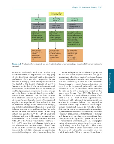
              Figure 23.2.  An	algorithm	for	the	diagnosis	and	basic	treatment	options	of	heartworm	disease	in	cats	in	which	heartworm	disease	is
              suspected.


              on  the  test  used  (Snyder  et  al.  2000).  Another  study,   Thoracic  radiography  and/or  echocardiography  are
              which evaluated Ab and Ag performance in a large group   the  two  most  useful  diagnostic  tests  after  serology  in
              of cats, also showed significant variation in diagnostic   feline patients exhibiting evidence of heartworm disease.
              performance  of  the  tests  when  compared  to  the  gold   Thoracic radiography is useful for diagnosis as well as
              standard of necropsy (which was imperfect because it   continued  monitoring  in  cases  of  feline  heartworm   Heartworm Disease
              was  limited  to  the  thorax)  (Berdoulay  et  al.  2004).   disease. The degree of caudal lobar arterial disease is best
              Because migrating L 4 and L 5 larvae and/or ectopic adult   assessed  by  the  dorsoventral  radiographic  projection
              worms would not have been detected by necropsy yet   (Nelson et al. 2005). The caudal lobar arteries, especially
              could still produce clinical signs and abnormal serologi-  the right, are the first to enlarge and typically are the
              cal results, the true number of infected cats was probably   most severely diseased (Figure 23.3). The diameters of
              underestimated.  Moreover,  cats  that  have  recovered   the caudal lobar arteries should not exceed that of the
              from heartworm infection may have persistent Ag and   ninth  rib  at  the  point  of  their  superimposition.
              Ab present for several months. In spite of these acknowl-  Cardiomegaly with right ventricular enlargement is less
              edged shortcomings, the study illustrated the limitations   common  in  heartworm-infected  cats  compared  to
              of heartworm serology in cats and that combining Ag   heartworm-infected  dogs.  Patchy  focal  or  diffuse  pul-
              and Ab tests results in improved detection of heartworm   monary  parenchymal  changes,  in  particular  a  bron-
              infection compared to using either test alone (but it also   chointerstitial pattern, are suggestive of (but not unique
              resulted  in  more  false-positive  results).  In  this  study,   to) feline heartworm disease. Less commonly associated
              antigen  tests  detected  79.3%  to  86.2%  of  heartworm   pulmonary findings include: hyperinflation of the lungs
              infections  and  were  highly  specific,  whereas  antibody   with  flattening  of  the  diaphragm,  consolidated  lung
              tests detected 62.1% to 72.4% of heartworm infections   lobes, pneumonitis (Figure 23.4), pleural effusion, and/
              with 1.4% to 19.1% false-positive results (compared to   or pneumothorax (Nelson et al. 2005). The radiographic
              0.3% to 2% for antigen tests). Because both L 5  larvae   picture often resembles feline asthma. However, radio-
              and adult worms are capable of causing clinical disease   graphic  changes  occur  less  frequently  in  feline  heart-
              in  the  cat,  both  antibody  and  antigen  tests  are  useful   worm  disease  than  in  canine  heartworm  disease  and
              tools, and the probability of making appropriate diag-  the  absence  of  radiographic  abnormalities  does  not
              nostic decisions improves when they are used together.  exclude a diagnosis of feline heartworm disease. In fact,

