Page 345 - Feline Cardiology
P. 345
358  Section K: Heartworm Disease


                                                                  A
                A


      Heartworm Disease                                           B


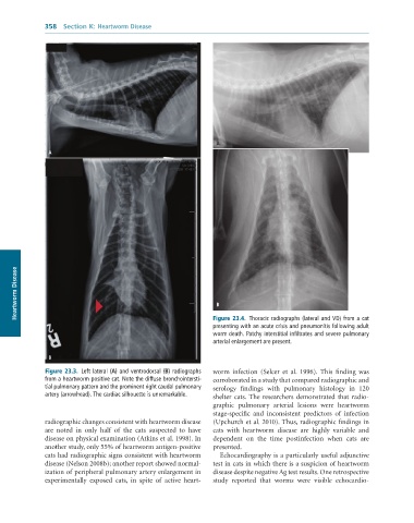
                                                                 Figure 23.4.  Thoracic	radiographs	(lateral	and	VD)	from	a	cat
                                                                 presenting	with	an	acute	crisis	and	pneumonitis	following	adult
                                                                 worm	death.	Patchy	interstitial	infiltrates	and	severe	pulmonary
                                                                 arterial	enlargement	are	present.

                B

               Figure 23.3.  Left	lateral	(A)	and	ventrodorsal	(B)	radiographs	  worm  infection  (Selcer  et  al.  1996).  This  finding  was
               from	a	heartworm	positive	cat.	Note	the	diffuse	bronchointersti-  corroborated in a study that compared radiographic and
               tial	pulmonary	pattern	and	the	prominent	right	caudal	pulmonary	  serology  findings  with  pulmonary  histology  in  120
               artery	(arrowhead).	The	cardiac	silhouette	is	unremarkable.  shelter  cats.  The  researchers  demonstrated  that  radio-
                                                                 graphic  pulmonary  arterial  lesions  were  heartworm
                                                                 stage-specific  and  inconsistent  predictors  of  infection
              radiographic changes consistent with heartworm disease   (Upchurch et al. 2010). Thus, radiographic findings in
              are  noted  in  only  half  of  the  cats  suspected  to  have   cats  with  heartworm  disease  are  highly  variable  and
              disease on physical examination (Atkins et al. 1998). In   dependent  on  the  time  postinfection  when  cats  are
              another study, only 55% of heartworm antigen-positive   presented.
              cats had radiographic signs consistent with heartworm   Echocardiography is a particularly useful adjunctive
              disease (Nelson 2008b); another report showed normal-  test in cats in which there is a suspicion of heartworm
              ization of peripheral pulmonary artery enlargement in   disease despite negative Ag test results. One retrospective
              experimentally  exposed  cats,  in  spite  of  active  heart-  study  reported  that  worms  were  visible  echocardio-

