Page 346 - Feline Cardiology
P. 346
Chapter 23: Heartworm Disease  359


              graphically in 40% of heartworm positive cats, including
              5 (of the 43 cats included) in which the Ag was negative
              (DeFrancesco  et  al.  2001).  However,  the  sensitivity  of
              echocardiography in these patients is highly operator-
              dependent and false positives are possible (Atwell et al.
              2001; Atkins et al. 2008). Two-dimensional echocardiog-                       RV
              raphy allows interrogation of the right heart, the main
              pulmonary  artery,  and  segments  of  the  right  and  left
              pulmonary arteries. Methodical examination is impor-                    LV
              tant since cats have light infections and detection may
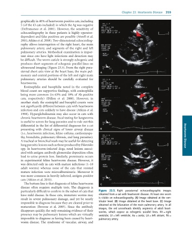
              be difficult. The worm cuticle is strongly echogenic and
              produces short segments of echogenic parallel lines on
              ultrasound imaging (Figure 23.5). From the right para-
              sternal short-axis view at the heart base, the main pul-  A
              monary and central portions of the left and right main
              pulmonary  arteries  should  be  carefully  evaluated  for
              heartworms.
                 Eosinophilia  and  basophilia  noted  in  the  complete
              blood count are supportive findings, with eosinophilia
              being more common (in 63% and 10% of Ab-positive
              cats,  respectively)  (Dillon  et  al.  2000).  However,  in                    AO
              another study the eosiniphil and baosphil counts were                                 PA
              not significantly different between cats with heartworm                   LA
              infection and cats unlikely to have disease (Atkins et al.
              1998). Hyperglobulinemia may also occur in cats with
              chronic heartworm disease. Fecal testing for lungworms
              is useful to screen for lung parasites and to rule out this
              differential in the list of differential diagnoses for a cat
              presenting  with  clinical  signs  of  lower  airway  disease   B
              (i.e., heartworm infection, feline-asthma, cardiomyopa-
              thy, bronchitis, pulmonary fibrosis, and lung parasites).                                                 Heartworm Disease
              A tracheal or bronchial wash may be useful for detecting
              lung parasitic lesions such as those produced by Filaroides
              spp.  In  heartworm-infected  dogs,  renal  lesions  associ-
              ated with antigen-antibody glomerular deposition often                          AO
              lead to urine protein loss. Similarly, proteinuria occurs
              in experimental feline heartworm disease. However, it                           PA
              was detected only in cats with mature infections (1–10
              adult  worms)  whereas  none  of  the  cats  that  resisted
              mature  infection  were  microalbuminuric.  Moreover  it
              was more common in heavily infected, antigen-positive
              cats (Atkins et al. 2010).
                 The bottom line is that diagnosis of feline heartworm   C
              disease  often  requires  multiple  tests.  The  diagnosis  is
              particularly difficult to confirm in the subset of cats that   Figure  23.5.  Right	 parasternal	 echocardiographic	 images
              have mild disease. In these cats, immature worms may   obtained	from	a	cat	with	heartworm	disease.	At	least	one	worm
              result in severe pulmonary damage, and yet be nearly   is	visible	on	echocardiography.	(A)	Image	obtained	at	the	ven-
              impossible to diagnose because they are cleared prior to   tricular	level.	(B)	Image	obtained	at	the	heart	base.	(C)	Image
              maturation  (Browne  et  al.  2005).  Since  the  antibody   obtained	at	the	bifurcation	of	the	main	pulmonary	artery.	In	all
                                                                 images,	 the	 red	 arrowheads	 identify	 segments	 of	 adult	 heart-
              disappears quickly, the only remaining evidence of their   worms,	 which	 appear	 as	 echogenic	 parallel	 lines.	 RV	=	right
              presence may be pulmonary lesions which are virtually   ventricle;	 LV	=	left	 ventricle;	 Ao	=	aorta;	 LA	=	left	 atrium;	 PA	=
              impossible to diagnose as having been caused by heart-  pulmonary	artery.
              worm  disease.  The  syndrome  of  vascular,  airway,  and

