
Page 1194 - Veterinary Toxicology, Basic and Clinical Principles, 3rd Edition
P. 1194
1126 SECTION | XVII Analytical Toxicology
VetBooks.ir pesticides, drugs of abuse, pesticide and drug metabolites, used. They often contain low concentrations of modifiers,
such as acetic or formic acid or buffers that contain phos-
mycotoxins, and marine toxins, such as domoic acid and
phate or acetate. These modifiers can greatly improve the
microcystins. HPLC is also used for the separation of
high molecular weight compounds such as proteins, peak shape and width for analytes and they may be crucial
although this type of analysis is not yet common in veteri- in providing good separation capabilities. Some of these
nary diagnostics. modifiers, particularly phosphate buffers, are not compati-
In its most basic form, an analytical HPLC system ble with the use of a mass spectrometer as the detector.
consists of a high pressure pump, a mobile phase, an
injector, a column, a detector, and the tubing that con-
nects these components. Modern commercially produced HPLC Detectors
systems typically include several other components. A Ultraviolet Light Detectors Ultraviolet spectrophoto-
degasser is used to remove dissolved gases such as nitro- metric (UV) detectors are commonly used in veterinary
gen and oxygen from the mobile phase as the presence of diagnostics. These detectors measure light absorbed by
these gases will result in inconsistent pressure in the sys- the mobile phase at different wavelengths. As compounds
tem. An autosampler is used to automatically inject sam- that absorb light at a specific wavelength elute, the absor-
ples and a temperature controlled column compartment is bance at that wavelength changes resulting in detection of
provided in order to provide a consistent column tempera- the compound. For single wavelength detectors, the wave-
ture. Of these different components, it is the column, the length of light must be selected as part of the analysis,
mobile phase, and the detector that vary most depending and only compounds that absorb light at that wavelength
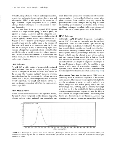
on the required analysis. will be detected. Variable wavelength detectors allow for
several different wavelengths or ranges of wavelengths to
HPLC Columns be used. Diode array detectors offer the capability to scan
across a wide range of wavelengths, producing a UV
As with GC, a wide variety of commercially produced
spectrum, which can be used in addition to the retention
HPLC columns allow for the analysis of many different
time to support the identification of an analyte (Fig. 81.7).
classes of analytes in different matrices. The sorbent in
the column (the “column packing”) typically provides
Fluorescence Detectors Another type of HPLC detector
separation based on the polarity of the analytes, although
commonly used in veterinary diagnostics is the fluores-
factors such as the pK a of the analyte may also be used to
cence detector. These detectors use high intensity light of a
provide separation. The length and diameter of the col-
specific wavelength to excite an analyte molecule to a
umn are also parameters that will affect the column’s sep-
higher energy state. The analyte quickly relaxes back to a
aration capabilities.
lower energy state, emitting light at a specific wavelength
as it does so. It is the emitted light that is detected and
HPLC Mobile Phases which provides the detector response. In order for this pro-
Mobile phases are chosen based on the separation needed, cess to work, the analyte must contain a functional group,
the type of column being used, and the detector. Typically, known as a fluorophore, which fluoresces. Aromatic ring
solvents such as water, methanol, and acetonitrile, are structures and carbonyl groups are among those that offer
FIGURE 81.7 An example of a spectrum taken from an ultraviolet light absorption detector. The x-axis is wavelength in nanometers and the y-axis
is intensity of detector response. The two distinctive features of this spectrum are the large peak at B235 nm and the broad, low intensity peak
centered at B325 nm.