Page 187 - Veterinary Toxicology, Basic and Clinical Principles, 3rd Edition
P. 187
154 SECTION | I General


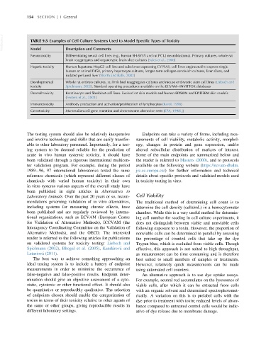
  VetBooks.ir  TABLE 9.5 Examples of Cell Culture Systems Used to Model Specific Types of Toxicity


               Model
                               Description and Comments
               Neurotoxicity   Differentiating neural cell lines (e.g., human SH-SY5Y and rat PC12 neuroblastoma). Primary cultures, whole rat
                               brain reaggregates and organotypic brain slice cultures (Sales et al., 2000)
               Hepatic toxicity  Human hepatoma HepG2 cell line and subclones expressing CYP1A1, cell lines engineered to express single
                               human or animal P450, primary hepatocyte cultures, longer-term collagen sandwich cultures, liver slices, and
                               isolated perfused liver (Worth and Balls, 2002)
               Developmental   Whole rat embryo cultures, rat limb bud reaggregates cultures and mouse embryonic stem cell lines (Liebsch and
               toxicity        Spielmann, 2002). Standard operating procedures available on the ECVAM INVITTOX databases
               Dermal toxicity  Keratinocyte and fibroblast cell lines. Excised rat skin models and human EPISKIN and EPIDERM skin models
                               (Fentem et al., 2001)
               Immunotoxicity  Antibody production and activation/proliferation of lymphocytes (Karol, 1998)
               Genotoxicity    Mammalian cell gene mutation and chromosome aberration tests (EPA, 1998i,j)


             The testing system should also be relatively inexpensive  Endpoints can take a variety of forms, including mea-
             and involve technology and skills that are easily transfer-  surements of cell viability, metabolic activity, morphol-
             able to other laboratory personnel. Importantly, for a test-  ogy, changes in protein and gene expression, and/or
             ing system to be deemed reliable for the prediction of  altered subcellular distribution of markers of interest.
             acute in vivo human systemic toxicity, it should have  Some of the main endpoints are summarized below and
             been validated through a rigorous international multicen-  the reader is referred to Masters (2000), and to protocols
             ter validation program. For example, during the period  available on the following website (https://ecvam-dbalm.
             1989 96, 97 international laboratories tested the same  jrc.ec.europa.eu/) for further information and technical
             reference chemicals (which represent different classes of  details about specific protocols and validated models used
             chemicals with varied human toxicity) in their own  in toxicity testing in vitro.
             in vitro systems various aspects of the overall study have
             been published in eight articles in Alternatives to
             Laboratory Animals. Over the past 20 years or so, recom-  Cell Viability
             mendations governing validation of in vitro alternatives,  The traditional method of determining cell count is to
             including systems for measuring chronic effects, have  determine the cell density (cells/mL) in a hemocytometer
             been published and are regularly reviewed by interna-  chamber. While this is a very useful method for determin-
             tional organizations, such as ECVAM (European Centre  ing cell number for seeding in cell culture experiments, it
             for Validation of Alternative Methods), ICCVAM (the  does not distinguish between viable and nonviable cells
             Interagency Coordinating Committee on the Validation of  following exposure to a toxin. However, the proportion of
             Alternative Methods), and the OECD. The interested  nonviable cells can be determined in parallel by assessing
             reader is referred to the following articles for publications  the percentage of counted cells that take up the dye
             on validated systems for toxicity testing: Liebsch and  Trypan blue, which is excluded from viable cells. Though
             Spielmann (2002), Bhogal et al. (2005), Kanda ´rova ´ and  effective, this approach is not suited to high throughput,
             Letasiova ´ (2011).                                as measurement can be time consuming and is therefore
                The best way to achieve something approaching an  best suited to small numbers of samples or treatments.
             ideal testing system is to include a battery of endpoint  However, relatively quick measurements can be made
             measurements in order to minimize the occurrence of  using automated cell counters.
             false-negative and false-positive results. Endpoint deter-  An alternative approach is to use dye uptake assays.
             mination should give an objective assessment of a cyto-  For example, neutral red accumulates on the lysosomes of
             static, cytotoxic or other functional effect. It should also  viable cells, after which it can be extracted from cells
             be quantitative or reproducibly qualitative. The selection  with an organic solvent and determined spectrophotomet-
             of endpoints chosen should enable the categorization of  rically. A variation on this is to prelabel cells with the
             toxins in terms of their toxicity relative to other agents of  dye prior to treatment with toxin; reduced levels of absor-
             the same or other groups, giving reproducible results in  bance compared to untreated control cells would be indic-
             different laboratory settings.                     ative of dye release due to membrane damage.

