
Page 281 - Veterinary Toxicology, Basic and Clinical Principles, 3rd Edition
P. 281
248 SECTION | II Organ Toxicity
VetBooks.ir distribution of lipidosis and inflammation is random and Steatosis Normal liver
multifocal. Conditions usually associated with steatohepatitis
are alcoholic liver disease, NAFLD, and endotoxemia sec-
ondary to intestinal disease. Any toxic compounds that can
cause steatosis can cause steatohepatitis if left untreated. ROS/RNS production
Ethanol
Steatohepatitis in humans can progress to fibrosis/cirrhosis
Endotoxemia
and hepatocellular carcinoma (Diehl, 2002). The clinical bio- ADH &
chemistry alterations include elevated serum transaminases. CYP2E1 Oxidative stress
Cytokines and
Acetaldehyde chemokine release
Hepatic Fibrosis
Fibrosis is a nonspecific lesion that usually results from Steatohepatitis Fibrosis/cirrhosis
chronic inflammation. Chronic inflammation can be the
result of continuous exposure to a variety of hepatotoxic
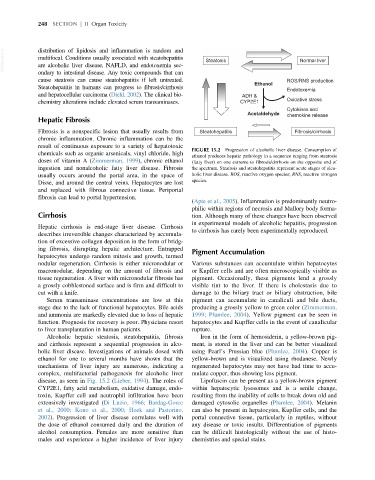
FIGURE 15.2 Progression of alcoholic liver disease. Consumption of
chemicals such as organic arsenicals, vinyl chloride, high
ethanol produces hepatic pathology in a sequence ranging from steatosis
doses of vitamin A (Zimmerman, 1999), chronic ethanol (fatty liver) on one extreme to fibrosis/cirrhosis on the opposite end of
ingestion and nonalcoholic fatty liver disease. Fibrosis the spectrum. Steatosis and steatohepatitis represent acute stages of alco-
usually occurs around the portal area, in the space of holic liver disease. ROS, reactive oxygen species; RNS, reactive nitrogen
Disse, and around the central veins. Hepatocytes are lost species.
and replaced with fibrous connective tissue. Periportal
fibrosis can lead to portal hypertension.
(Apte et al., 2005). Inflammation is predominantly neutro-
philic within regions of necrosis and Mallory body forma-
Cirrhosis tion. Although many of these changes have been observed
in experimental models of alcoholic hepatitis, progression
Hepatic cirrhosis is end-stage liver disease. Cirrhosis
to cirrhosis has rarely been experimentally reproduced.
describes irreversible changes characterized by accumula-
tion of excessive collagen deposition in the form of bridg-
ing fibrosis, disrupting hepatic architecture. Entrapped Pigment Accumulation
hepatocytes undergo random mitosis and growth, termed
nodular regeneration. Cirrhosis is either micronodular or Various substances can accumulate within hepatocytes
macronodular, depending on the amount of fibrosis and or Kupffer cells and are often microscopically visible as
tissue regeneration. A liver with micronodular fibrosis has pigment. Occasionally, these pigments lend a grossly
a grossly cobblestoned surface and is firm and difficult to visible tint to the liver. If there is cholestasis due to
cut with a knife. damage to the biliary tract or biliary obstruction, bile
Serum transaminase concentrations are low at this pigment can accumulate in canaliculi and bile ducts,
stage due to the lack of functional hepatocytes. Bile acids producing a grossly yellow to green color (Zimmerman,
and ammonia are markedly elevated due to loss of hepatic 1999; Plumlee, 2004). Yellow pigment can be seen in
function. Prognosis for recovery is poor. Physicians resort hepatocytes and Kupffer cells in the event of canalicular
to liver transplantation in human patients. rupture.
Alcoholic hepatic steatosis, steatohepatitis, fibrosis Iron in the form of hemosiderin, a yellow-brown pig-
and cirrhosis represent a sequential progression in alco- ment, is stored in the liver and can be better visualized
holic liver disease. Investigations of animals dosed with using Pearl’s Prussian blue (Plumlee, 2004). Copper is
ethanol for one to several months have shown that the yellow-brown and is visualized using rhodanese. Newly
mechanisms of liver injury are numerous, indicating a regenerated hepatocytes may not have had time to accu-
complex, multifactorial pathogenesis for alcoholic liver mulate copper, thus showing less pigment.
disease, as seen in Fig. 15.2 (Lieber, 1994). The roles of Lipofuscin can be present as a yellow-brown pigment
CYP2E1, fatty acid metabolism, oxidative damage, endo- within hepatocytic lysosomes and is a senile change,
toxin, Kupffer cell and neutrophil infiltration have been resulting from the inability of cells to break down old and
extensively investigated (Di Luzio, 1966; Bardag-Gorce damaged cytosolic organelles (Plumlee, 2004). Melanin
et al., 2000; Kono et al., 2000; Hoek and Pastorino, can also be present in hepatocytes, Kupffer cells, and the
2002). Progression of liver disease correlates well with portal connective tissue, particularly in reptiles, without
the dose of ethanol consumed daily and the duration of any disease or toxic insults. Differentiation of pigments
alcohol consumption. Females are more sensitive than can be difficult histologically without the use of histo-
males and experience a higher incidence of liver injury chemistries and special stains.