
Page 284 - Veterinary Toxicology, Basic and Clinical Principles, 3rd Edition
P. 284
Liver Toxicity Chapter | 15 251
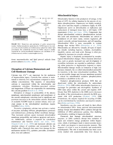
VetBooks.ir Cl Cl Cl CYP2E1 Cl C Cl Cl Mitochondrial Injury
C
Mitochondria function in the production of energy, in the
Cl
Cl
dative phosphorylation. Hepatocytes are highly metaboli-
Carbon Covalent binding Trichlorocarbon form of ATP, for cellular function by the process of oxi-
tetrachloride radical cally active and thus require a continuous supply of ATP.
O 2 Hepatocytes active in detoxification or replication and
Lipid peroxidation replacement of damaged tissue have a still higher ATP
O
requirement (Dahm and Jones, 1996). Compounds that
Covalent binding
Cl C Cl disrupt mitochondrial oxidative phosphorylation include
Hepatic necrosis Cl bile acids and amiodarone. Mitochondria are critical to
(centrilobular) Trichloroperoxy modulation of cell redox status, osmotic regulation, pH
radical
control, cytosolic calcium homeostasis, and cell signaling.
Mitochondrial DNA is more susceptible to oxidant
FIGURE 15.5 Metabolism and mechanism of carbon tetracholoride
toxicity. Carbon tetrachloride metabolism by CYP450 leads to free radi- damage than nuclear DNA (Stirnimann et al., 2010).
cals such as trichlorocarbon and trichloroperoxy radical that initiate lipid Mitochondria are affected by virtually all types of injuri-
peroxidation. The centrilobular location of CYP2E1 enzyme is mainly ous stimuli, including hypoxia, oxidants, electrophiles,
responsible for carbon tetrachloride metabolism and contributes to cen- lipophilic cations, and weak acids. Damage is often pre-
trilobular necrosis similar to acetaminophen toxicity.
cipitated by increases in cytosolic calcium.
Hepatic injury is frequently accompanied by morpho-
logical mitochondrial changes. These structural abnormal-
tissue macromolecules and lipid peroxyl radicals form
ities, such as greatly increased size and development of
protein adducts (Jaeschke, 2008).
crystalline inclusions, are regarded as pathologic, reflect-
ing either protective or degenerative response to injury.
Mitochondrial damage results in formation of high con-
Disruption of Calcium Homeostasis and ductance channels, the so-called mitochondrial permeabil-
Cell Membrane Damage ity transition, in the inner mitochondrial membrane. This
21 is an irreversible change and, because membrane potential
Calcium ions (Ca ) are important for the mediation
is critical for mitochondrial oxidative phosphorylation,
of hepatocellular injury. Cytosolic-free calcium is main-
constitutes a deathblow to the cell.
tained at relatively low concentrations compared to extra-
Oxidative phosphorylation produces ROS that are
cellular concentrations. The majority of intracellular
deactivated within the mitochondria by antioxidants
calcium is sequestered within the mitochondria and
(Watkins, 1999). GSH within mitochondria functions as a
endoplasmic reticulum. Membrane-associated calcium
scavenger for peroxides and electrophiles. Synthesis of
and magnesium ATPases are responsible for maintaining
GSH takes place within the cytosol and requires ATP.
this calcium gradient (Farrell et al., 1990).
Greater than 90% depletion in the GSH reserve decreases
Disruption or changes in permeability of the plasma
the ability of the mitochondrion to detoxify the ROS asso-
membrane, mitochondrial membranes and membranes of
ciated with ATP production (Watkins, 1999). GSH
the smooth endoplasmic reticulum lead to significant and
S-transferase, the enzyme required for recycling of GSH,
persistent increases in the intracellular calcium. Depletion
can be overwhelmed by xenobiotics and reactive metabo-
of available NADPH leads to calcium release, since cal-
lites (Dahm and Jones, 1996).
cium pumps in the mitochondrial membrane require
Xenobiotics can also cause cell death by their effects
NADPH (Cullen, 2005).
on mitochondrial DNA. Some antiviral deoxynucleoside
Excessive cytoplasmic calcium ions activate a variety
analogs disrupt mitochondrial DNA synthesis through the
of enzymes, including ATPases, phospholipases, proteases
inhibition of DNA polymerase gamma, thus depleting
and endonucleases, producing further membrane damage.
mitochondria, resulting in hepatocyte death.
Thus increased calcium causes increased mitochondrial
Chemicals that damage mitochondrial structure,
membrane permeability and induces apoptosis and necro-
enzymes, or DNA synthesis disrupt beta oxidation of
sis. Additionally, calcium is required for cytoskeletal
lipids and oxidative energy production within hepato-
maintenance and function (Dahm and Jones, 1996;
cytes, which, if prolonged, leads to microvesicular steato-
Delgado-Coello et al., 2006) and increased calcium can
sis, which can progress to macrovesicular steatosis. This
stimulate release of cytokines and eicosanoids by the
sequence of events is seen in alcoholic and nonalcoholic
Kupffer cells. Chemicals that cause liver damage by this
steatohepatitis. The role of mitochondria has been exten-
mechanism include CCl 4 , quinines, peroxides, acetamino-
sively studied with nonalcoholic fatty liver disease in
phen, iron and cadmium.