
Page 282 - Veterinary Toxicology, Basic and Clinical Principles, 3rd Edition
P. 282
Liver Toxicity Chapter | 15 249
VetBooks.ir Hepatic Neoplasia by hepatic fibrosis as well as biliary hyperplasia and
sometimes nodular regeneration of parenchyma.
Chemically induced neoplasms can originate from hepato-
cytes, biliary epithelium, and very rarely from sinusoidal
endothelium. Neoplasms occur months or years after toxin MECHANISMS OF LIVER DAMAGE
exposure (Plumlee, 2004). If a toxin produces direct dam-
Mechanisms of liver injury have been classified as either
age to DNA, a single exposure can lead eventually to neo-
intrinsic or idiosyncratic. Intrinsic injury can produce
plastic changes (Zimmerman, 1999; Pineiro-Carrero and
steatosis, necrosis, cholestasis, or multiple lesions, often
Pineiro, 2004). Aflatoxin B 1 is an agent that acts by
with minimal inflammation (Sturgill and Lambert, 1997).
alkylating DNA and is associated with hepatocellular
Intrinsic liver injury is a predictable, reproducible, dose-
carcinoma in humans infected with hepatitis B virus.
dependent response to a xenobiotic (Dahm and Jones,
Aflatoxin B 1 has also been implicated in liver cancers in
1996; Sturgill and Lambert, 1997; Zimmerman, 1999;
trout and laboratory animals (Newberne and Butler,
Pineiro-Carrero and Pineiro, 2004). A threshold dose
1969). Nongenotoxic agents must be given at high doses
exists for xenobiotics causing intrinsic liver injury, and
for long periods of time to induce cancer. Examples of
there is often a predictable latent period between the time
nongenotoxic agents include phthalate esters in plastici-
of exposure and clinical evidence of liver injury. Intrinsic
zers, phenoxy acid herbicides and hypolipidimic drugs.
liver injury accounts for the vast majority of toxic liver
Hepatic adenomata are benign hepatocytic tumors
injury cases and is often produced by reactive products
associated with contraceptive steroid use in humans
of xenobiotic metabolism, such as electrophiles or free
(Zimmerman, 1999). Hepatocellular carcinomata are
radicals, though a few drugs cause intrinsic liver injury
malignant neoplasms induced by various chemicals and
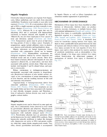
without bioactivation. An abbreviated summary of
botanical toxins and are of more concern. These have
mechanisms of intrinsic liver injury is illustrated in
been found in humans affected with hepatitis B virus and
Fig. 15.3.
exposed to aflatoxin B 1 , as noted above, and have been
associated with anabolic steroid abuse. Other risk factors
Excretion
include viral hepatitis C and D, ethanol abuse, and expo-
Phase III
sure to microcystin. transporters
Biliary carcinomata or cholangiocarcinomata are
Inactive
uncommonly associated with exposure to drugs/chemi- Cholestasis
Metabolite
cals. Biochemical indicators of note include normal, ele-
vated, or low concentrations of serum transaminases, loss Phase II Metabolism
of liver function, and the resulting increase in bile acid
and ammonia concentrations. Bile
Phase I Metabolism
Angiosarcomata or hemangiosarcomata derive from
Direct
sinusoidal epithelium (Zimmerman, 1999; Treinen- Xenobiotic
Damage
Moslen, 2001; Plumlee, 2004). These neoplasms are rare
Conjugation
but rapidly lethal and have been associated with exposure
Phase I Metabolism
to vinyl chloride, inorganic arsenics, and Thorotrast, a
form of radioactive thorium dioxide once used in radio- Phase II
Active Metabolism
graphic contrast studies.
Metabolite
Megalocytosis DNA
Damage Electrophile Free Radical
Hepatic megalocytosis can be observed in some aged ani-
mals, but is characteristic lesion observed in toxic insult
by pyrrolizidine alkaloids and aflatoxins. Megalocytosis Carcinogenesis
Protein Membrane
is caused by impaired cell division, resulting in enlarged Damage Peroxidation
hepatocytes with markedly enlarged nuclei (Plumlee,
2004). Pyrrolizidine alkaloids have an antimitotic effect FIGURE 15.3 Basic mechanisms of hepatic injury showing the rela-
on the hepatocytes but don’t inhibit DNA synthesis. Since tionship between multiple pathways for metabolism and toxicity for any
hepatocytes naturally proliferate to replace the damaged compound. The liver metabolizes xenobiotics (and some endogenous
compounds) to form water-soluble products appropriate for urinary or
cells, hepatocytes are active in DNA and protein synthe-
biliary excretion. Some compounds are activated through these metabolic
sis; when they cannot divide, megalocytosis results. Very processes to free radicals, electrophiles or other toxic products that may
often, chronic pyrrolizidine intoxication is accompanied induce hepatic injury.