
Page 320 - Veterinary Toxicology, Basic and Clinical Principles, 3rd Edition
P. 320
Reproductive Toxicity and Endocrine Disruption Chapter | 17 287
VetBooks.ir and/or placental origin, exposure to exogenous HAAs classified as primordial, primary (some become atretic),
secondary and tertiary (antral) follicles based on their
and seasonal influences (Ginther, 1992; Senger, 2003;
stage of development (Evans et al., 2007; Evans and
Evans et al., 2007).
The primary functions of the ovary are oogenesis (pro- Ganjam, 2017).
duction of female gametes (oocytes or ova)) and steroido- A primary oocyte surrounded by a single, flattened
genesis (production of estrogens and progesterone). The cell layer is a primordial follicle. A basal lamina sepa-
ovaries of most domestic mammals consist of a peripheral rates the single layer of what will become granulosa cells
parenchymatous zone (cortex), containing various stages from the adjacent stromal tissue which eventually devel-
of follicular and luteal gland development and a central ops into the theca cells (theca interna and theca externa).
vascular zone (medulla), comprised of collagenous con- The granulosa cells homologous to the Sertoli cells in the
nective tissue rich in blood vessels (Senger, 2003; Evans testis, and the theca interna cells are the female equiva-
and Ganjam, 2017)(Figure 17.3). The structural and func- lent of the Leydig cells (Senger, 2005). Following the
tional unit of the ovary is the follicle. Follicles are appropriate endocrine stimulation, primordial follicles
are recruited to undergo possible further differentiation
into estrogen-producing antral follicles and ultimately
ovulation, which results in the release of a secondary
oocyte (primary oocyte in dogs) and formation of a cor-
pus luteum (CL) which produces progesterone (Ginther,
1992; Senger, 2003; Evans et al., 2007; Evans and
Ganjam, 2017).
Female Reproductive Physiology
Females are born with a finite pool of primordial follicles
(up to hundreds of thousands), and reproductive cyclicity
(i.e., estrous or menstrual cycles) provides females with
repeated opportunities for the establishment of pregnancy.
The majority of mammalian species (subprimates) have
estrous cycles, which reflect the physiological changes
occurring between successive ovulations and/or periods of
sexual receptivity (estrus) (Senger, 2005; Evans and
Ganjam, 2017). Humans and non-human primates experi-
ence menstrual rather than estrous cycles and do not have
defined periods of sexual receptivity (i.e., estrus). Unlike
the estrous cycles in subprimates, the reproductive cycle
in menstruating animals is divided into phases (i.e., men-
ses, proliferative and secretory phases), which are defined
based on the physiological state of the uterine endome-
trium, rather than on the predominant ovarian structures
(Senger, 2003; Genuth, 2004b; Evans and Ganjam, 2017).
The Estrous Cycle
The follicular and luteal phases of the estrous cycle
describe the predominant ovarian structures and the cor-
responding gonadal steroid concentrations which result
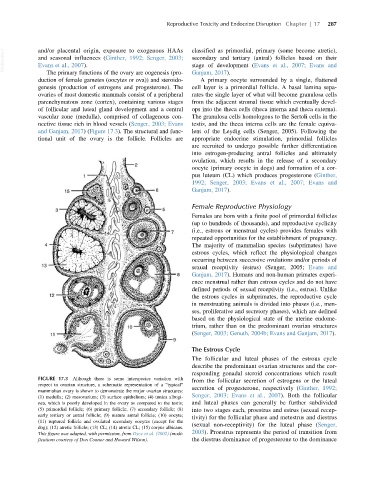
FIGURE 17.3 Although there is some interspecies variation with
from the follicular secretion of estrogens or the luteal
respect to ovarian structure, a schematic representation of a “typical”
secretion of progesterone, respectively (Ginther, 1992;
mammalian ovary is shown to demonstrate the major ovarian structures:
(1) medulla; (2) mesovarium; (3) surface epithelium; (4) tunica albugi- Senger, 2003; Evans et al., 2007). Both the follicular
nea, which is poorly developed in the ovary as compared to the testis; and luteal phases can generally be further subdivided
(5) primordial follicle; (6) primary follicle; (7) secondary follicle; (8) into two stages each, proestrus and estrus (sexual recep-
early tertiary or antral follicle; (9) mature antral follicle; (10) oocyte; tivity) for the follicular phase and metestrus and diestrus
(11) ruptured follicle and ovulated secondary oocytes (except for the
dog); (12) atretic follicle; (13) CL; (14) atretic CL; (15) corpus albicans. (sexual non-receptivity) for the luteal phase (Senger,
This figure was adapted, with permission, from Dyce et al. (2002) (modi- 2003). Proestrus represents the period of transition from
fications courtesy of Don Connor and Howard Wilson). the diestrus dominance of progesterone to the dominance