
Page 315 - Veterinary Toxicology, Basic and Clinical Principles, 3rd Edition
P. 315
282 SECTION | II Organ Toxicity
VetBooks.ir ejaculation of sperm. The primary functions of the testis Senger, 2003; Evans and Ganjam, 2017). In some species,
the rete tubules coalesce in a fibrous region of the testis
(testicle) are spermatogenesis (production of male
referred to as the mediastinum, which joins with septal
gametes (sperm or spermatozoa)) and steroidogenesis
(production of androgens and estrogens). Unlike the projections of the tunica albuginea, part of the testicular
female in which oogonia are no longer replicating and the capsule (Senger, 2003). The rete tubules join with the
full complement of potential oocytes are present at birth, efferent ductules which attach to the epididymidis.
spermatogonia are proliferating and differentiating into Within the seminiferous tubules are germ cells at var-
spermatozoa continuously, and the testis is organized in ious stages of differentiation and Sertoli cells which pro-
such a way as to maximize sperm production (Senger, vide germ cells with structural support and nutrients, as
2003; Evans and Ganjam, 2017). well as regulatory and paracrine factors (Thomas and
Thomas, 2001)(Figure 17.1B). Tight junctions (junc-
tional complexes) between adjacent Sertoli cells divide
Testicular Structure the seminiferous epithelium into basal and adluminal
The parenchyma of the testis is divided into the tubular compartments, with Sertoli cells anchored to the base-
and interstitial compartments (Senger, 2003; Evans and ment membrane and surrounding the developing popula-
Ganjam, 2017)(Figure 17.1A). The structural and func- tions of germ cells (Thomas and Thomas, 2001; Genuth,
tional units of the testis are the seminiferous tubules 2004b; Senger, 2003; Evans and Ganjam, 2017). The
within the tubular compartment and the Leydig (intersti- seminiferous tubules are surrounded by peritubular myoid
tial cells) within the interstitial compartment. Depending cells, which in combination with the junctional com-
on the species, it is estimated that the seminiferous plexes, form the “blood testis barrier” to prevent free
tubules comprise approximately 80% of the adult testis, exchange of large proteins and some xenobiotics between
with the interstitium comprising most of the remaining the blood and the fluid within the seminiferous tubules
20% (Genuth, 2004b). Seminiferous tubules form highly (Thomas and Thomas, 2001; Senger, 2003; Evans and
convoluted loops (tubulus contortus) which begin and end Ganjam, 2017).
with straight portions (tubulus rectus) that connect to the Within the interstitial compartment are the Leydig
rete tubules (Thomas and Thomas, 2001; Genuth, 2004b; (interstitial) cells, as well as capillaries, lymphatic vessels
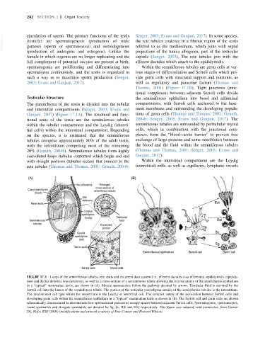
FIGURE 17.1 Loops of the seminiferous tubules, rete testis and excurrent duct system (i.e., efferent ductules (vas efferentia), epididymidis (epididy-
mis) and ductus deferens (vas deferens)), as well as a cross-section of a seminiferous tubule showing the microanatomy of the seminiferous epithelium
in a “typical” mammalian testis, are shown in (A). Mature spermatozoa follow the pathway denoted by arrows. Testicular fluid is secreted by the
Sertoli cell into the lumen of the seminiferous tubule. The portion of the testicular parenchyma outside of the seminiferous tubules is the interstitium.
The predominant cell type within the interstitium is the Leydig or interstitial cell. The complex nature of the association between Sertoli cells and
developing germ cells within the seminiferous epithelium in a “typical” mammalian testis is shown in (B). The Sertoli cell and germ cells are shown
schematically disassociated to demonstrate how spermatozoal precursors occupy spaces between adjacent Sertoli cells. Spermatogonia, spermatocytes,
round spermatids and elongate spermatids are denoted by Sg, Sc, RSt and ESt, respectively. This figure was adapted, with permission, from Garner
DL, Hafez ESE (2000) (modifications and artwork courtesy of Don Connor and Howard Wilson).