
Page 318 - Veterinary Toxicology, Basic and Clinical Principles, 3rd Edition
P. 318
Reproductive Toxicity and Endocrine Disruption Chapter | 17 285
VetBooks.ir Haschek et al., 2010). The number and duration of the var- Endocrine Regulation of Spermatogenesis
ious stages of the cycle of the somniferous epithelium
While the female hypothalamus has both fully developed
species
2003),
and
with
various
(Senger,
vary
classification schemes have been used, based on the mor- tonic and surge centers for GnRH release (especially prior
to ovulation), the hypothalamic GnRH surge center in the
phological characteristics of the spermatid nucleus or the male is diminished, and the anterior pituitary gland of the
development of the acrosomic system (Franca et al., male does not experience surges in GnRH stimulation
¸
2005). In subprimates, sequential stages are arranged (Senger, 2003; Evans and Ganjam, 2017). This gender-
along the length of the seminiferous tubule in consecutive specific alteration in the hypothalamus facilitates the
order, forming a “spermatogenic wave” (Senger, 2003; normal endocrine milieu which maintains continuous
Haschek et al., 2010). The progeny of one spermatogo- spermatogenesis and stimulates normal sexual behavior
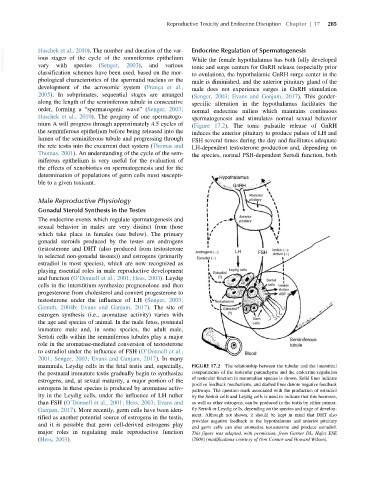
nium A will progress through approximately 4.5 cycles of (Figure 17.2). The tonic pulsatile release of GnRH
the seminiferous epithelium before being released into the induces the anterior pituitary to produce pulses of LH and
lumen of the seminiferous tubule and progressing through FSH several times during the day and facilitates adequate
the rete testis into the excurrent duct system (Thomas and LH-dependent testosterone production and, depending on
Thomas, 2001). An understanding of the cycle of the sem- the species, normal FSH-dependent Sertoli function, both
iniferous epithelium is very useful for the evaluation of
the effects of xenobiotics on spermatogenesis and for the
determination of populations of germ cells most suscepti-
ble to a given toxicant.
Male Reproductive Physiology
Gonadal Steroid Synthesis in the Testes
The endocrine events which regulate spermatogenesis and
sexual behavior in males are very distinct from those
which take place in females (see below). The primary
gonadal steroids produced by the testes are androgens
(testosterone and DHT (also produced from testosterone
in selected non-gonadal tissues)) and estrogens (primarily
estradiol in most species), which are now recognized as
playing essential roles in male reproductive development
and function (O’Donnell et al., 2001; Hess, 2003). Leydig
cells in the interstitium synthesize pregnenolone and then
progesterone from cholesterol and convert progesterone to
testosterone under the influence of LH (Senger, 2003;
Genuth, 2004b; Evans and Ganjam, 2017). The site of
estrogen synthesis (i.e., aromatase activity) varies with
the age and species of animal. In the male fetus, postnatal
immature male and, in some species, the adult male,
Sertoli cells within the seminiferous tubules play a major
role in the aromatase-mediated conversion of testosterone
to estradiol under the influence of FSH (O’Donnell et al.,
2001; Senger, 2003; Evans and Ganjam, 2017). In many
mammals, Leydig cells in the fetal testis and, especially, FIGURE 17.2 The relationship between the tubular and the interstitial
the postnatal immature testis gradually begin to synthesize compartments of the testicular parenchyma and the endocrine regulation
of testicular function in mammalian species is shown. Solid lines indicate
estrogens, and, at sexual maturity, a major portion of the
positive feedback mechanisms, and dashed lines denote negative feedback
estrogens in these species is produced by aromatase activ-
pathways. The question mark associated with the production of estradiol
ity in the Leydig cells, under the influence of LH rather by the Sertoli cells and Leydig cells is used to indicate that this hormone,
than FSH (O’Donnell et al., 2001; Hess, 2003; Evans and as well as other estrogens, can be produced in the testis by either primar-
Ganjam, 2017). More recently, germ cells have been iden- ily Sertoli or Leydig cells, depending on the species and stage of develop-
ment. Although not shown, it should be kept in mind that DHT also
tified as another potential source of estrogens in the testis,
provides negative feedback to the hypothalamus and anterior pituitary
and it is possible that germ cell-derived estrogens play
and germ cells can also aromatize testosterone and produce estradiol.
major roles in regulating male reproductive function This figure was adapted, with permission, from Garner DL, Hafez ESE
(Hess, 2003). (2000) (modifications courtesy of Don Connor and Howard Wilson).