
Page 327 - Veterinary Toxicology, Basic and Clinical Principles, 3rd Edition
P. 327
294 SECTION | II Organ Toxicity
VetBooks.ir
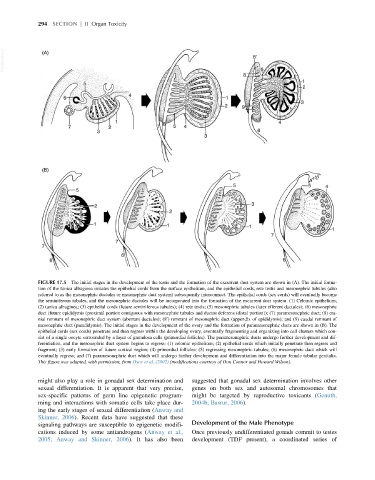
FIGURE 17.5 The initial stages in the development of the testis and the formation of the excurrent duct system are shown in (A). The initial forma-
tion of the tunica albuginea isolates the epithelial cords from the surface epithelium, and the epithelial cords, rete testis and mesonephric tubules (also
referred to as the mesonephric ductules or mesonephric duct system) subsequently interconnect. The epithelial cords (sex cords) will eventually become
the seminiferous tubules, and the mesonephric ductules will be incorporated into the formation of the excurrent duct system. (1) Celomic epithelium;
(2) tunica albuginea; (3) epithelial cords (future seminiferous tubules); (4) rete testis; (5) mesonephric tubules (later efferent ductules); (6) mesonephric
duct (future epididymis (proximal portion contiguous with mesonephric tubules and ductus deferens (distal portion)); (7) paramesonephric duct; (8) cra-
nial remnant of mesonephric duct system (aberrant ductules); (8 ) remnant of mesonephric duct (appendix of epididymis); and (9) caudal remnant of
0
mesonephric duct (paradidymis). The initial stages in the development of the ovary and the formation of paramesonephric ducts are shown in (B). The
epithelial cords (sex cords) penetrate and then regress within the developing ovary, eventually fragmenting and organizing into cell clusters which con-
sist of a single oocyte surrounded by a layer of granulosa cells (primordial follicles). The paramesonephric ducts undergo further development and dif-
ferentiation, and the mesonephric duct system begins to regress: (1) celomic epithelium; (2) epithelial cords which initially penetrate then regress and
fragment; (3) early formation of future cortical region; (4) primordial follicles; (5) regressing mesonephric tubules; (6) mesonephric duct which will
eventually regress; and (7) paramesonephric duct which will undergo further development and differentiation into the major female tubular genitalia.
This figure was adapted, with permission, from Dyce et al. (2002) (modifications courtesy of Don Connor and Howard Wilson).
might also play a role in gonadal sex determination and suggested that gonadal sex determination involves other
sexual differentiation. It is apparent that very precise, genes on both sex and autosomal chromosomes that
sex-specific patterns of germ line epigenetic program- might be targeted by reproductive toxicants (Genuth,
ming and interactions with somatic cells take place dur- 2004b; Basrur, 2006).
ing the early stages of sexual differentiation (Anway and
Skinner, 2006). Recent data have suggested that these
signaling pathways are susceptible to epigenetic modifi- Development of the Male Phenotype
cations induced by some antiandrogens (Anway et al., Once previously undifferentiated gonads commit to testes
2005; Anway and Skinner, 2006). It has also been development (TDF present), a coordinated series of