
Page 329 - Veterinary Toxicology, Basic and Clinical Principles, 3rd Edition
P. 329
296 SECTION | II Organ Toxicity
VetBooks.ir caused by fetal movements and myometrial contractions, THE MECHANISMS AND EFFECTS
Normal parturition approaches as neural signals
OF REPRODUCTIVE TOXICANTS
along with elevated basal levels of oxytocin and increased
secretion of PGF 2α , bring about the first stage of labor. A The Effects of EDCs on Wildlife Species,
rapid increase in oxytocin and PGF 2α secretion leads to Humans and Domestic Animals
rupture of the allantochorionic membrane and the com-
It should be evident from the previous discussion that
mencement of the second stage of labor. Strong myome-
maximum reproductive efficiency, including normal
trial contractions result in the delivery of offspring, as
embryonic and fetal development, is dependent on the
well as the expulsion of the fetal membranes during the
structural and functional integrity of multiple organs
third stage of labor (Senger, 2003; Evans et al., 2007).
and tissues, as well as various signaling pathways
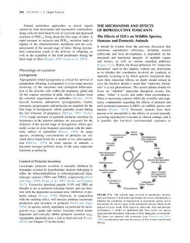
(Figure 17.6). Within the broad definition for “endocrine
disruption” used in this chapter, without any restrictions
Physiology of Lactation
as to whether the xenobiotics involved are synthetic or
Lactogenesis naturally occurring or by which specific mechanism they
Appropriately timed lactogenesis is critical for survival of exert their endocrine effects, no doubt should remain in
mammalian offspring. Lactogenesis is a two-stage process even the harshest skeptic’s mind that “endocrine disrup-
involving: (1) the enzymatic and cytological differentia- tion” is a real phenomenon. The current debate should not
tion of the alveolar cells within the mammary gland and focus on “whether” endocrine disruption occurs, but,
(2) the copious secretion of milk, which is distinct from rather, “when” it occurs and under what circumstances.
the colostral sequestration of antibodies (Tucker, 1994). There is increasing concern within the scientific and regu-
Growth hormone, aldosterone, prostaglandins, insulin, latory communities regarding the effects of prenatal and
estrogens, progestagens and prolactin are required for the early postnatal exposures to EDCs on wildlife species and
first stage of lactogenesis, which generally occurs during humans (Evans, 2017). Domestic animals have been
the last trimester of pregnancy (McCue, 1993; Tucker, known to be susceptible to the effects of several naturally
1994). Large increases in pulsatile prolactin secretion by occurring reproductive toxicants in clinical settings, and it
lactotropes in the anterior pituitary are necessary for the is possible that low-level environmental exposures to
initiation of the second stage of lactogenesis, which gen-
erally occurs in close temporal association with the endo-
crine milieu of parturition (Evans, 1996). In many
species, circulating concentrations of prolactin are ele-
vated above basal levels for a month or two after parturi-
tion (McCue, 1993). In some species of animals, a
placental lactogen performs many of the same endocrine
functions as prolactin.
Control of Prolactin Secretion
Lactotropic prolactin secretion is tonically inhibited by
dopamine secreted by hypothalamic neurons belonging to
either the tuberoinfundibular or tuberohypophysial dopa-
minergic systems (TIDA and THDA, respectively) (Neill
and Nagy, 1994; Evans et al., 2007; Evans and Ganjam,
2017). Vasoactive intestinal peptide (VIP) and TRH are
thought to act as prolactin releasing factors and can inter-
fere with the dopamine-associated tonic inhibition of pro-
lactin release (Evans, 1996). Oxytocin, in conjunction FIGURE 17.6 The multiple steps involved in reproductive develop-
ment and function in both males and females are shown schematically to
with the suckling reflex, will increase pituitary lactotropic
illustrate the complexity of reproduction in mammalian species and to
production and secretion of prolactin (Neill and Nagy, demonstrate the various stages in the reproductive process which can be
1994). In species strictly dependent on prolactin for lacto- targeted for toxic insult. With respect to embryonic, fetal and placental
genesis (e.g., equids and swine), toxicants which mimic development, it should be understood that these events are species
dopamine and tonically inhibit prolactin secretion (e.g., dependent and interrelated, with many of them taking place concurrently.
This figure was adapted, with permission, from Ellington and Wilker
ergopeptine alkaloids) pose a risk to fetal survival (Evans,
(2006) (modifications and artwork courtesy of Don Connor and Howard
2011b; see Chapter 72 in this book). Wilson).