
Page 720 - Veterinary Toxicology, Basic and Clinical Principles, 3rd Edition
P. 720
PCBs, PBBs, Dioxin, PCDDs Chapter | 51 685
VetBooks.ir
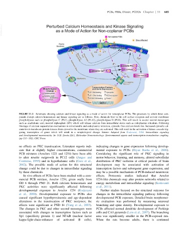
FIGURE 51.3 Schematic showing calcium and kinase signaling as a mode of action for noncoplanar PCBs. The processes by which these com-
pounds disrupt calcium homeostasis and kinase signaling are as follows. First, chemicals bind to the cell surface receptors and activate membrane
phospholipases such as phospholipase C (PLC), phospholipase A2 (PLA2), phospholipase D (PLD). This will result in several second messengers
such as arachidonic acid, inositol triphosphate (IP3) which will release calcium from intracellular stores such as endoplasmic reticulum. Following
blockage of calcium sequestration mechanisms in mitochondria and endoplasmic reticulum, cytosolic free calcium levels rise. Increased cytosolic cal-
cium levels translocate protein kinases from cytosol to the membrane where they are activated. This will result in the activation of kinase cascade trig-
gering transcription of genes which will result in a morphological change. Source: Adapted from Kodavanti, 2004. Intracellular signaling
and developmental neurotoxicity. In: N.H. Zawia (Ed.), Molecular Neurotoxicology: Environmental agents and transcription-transduction coupling.
(pp 151 182). CRC Press.
no effects on PKC translocation. Literature reports indi- indicating changes in gene expression following develop-
cate that at slightly higher concentrations, commercial mental exposure to PCBs (Riyaz Basha et al., 2006).
PCB mixtures (Aroclors 1221 and 1254) have been able Considering the significant role of PKC signaling in
to alter neurite outgrowth in PC12 cells (Angus and motor behavior, learning, and memory, altered subcellular
Contreras, 1995) and in hypothalamic cells (Gore et al., distribution of PKC isoforms at critical periods of brain
2002). The possible mode of action for this structural development may be associated with activation of
change could be due to changes in intracellular signaling transcription factors and subsequent gene expression, and
by these chemicals. may be a possible mechanism of PCB-induced neurotoxic
In vivo effects of PCBs have been studied with a com- effects. Proteomic studies indicated that Aroclor
mercial PCB mixture, Aroclor 1254, given orally from 1254-like chemicals may alter protein networks related to
GD 6 through PND 21. Both calcium homeostasis and energy metabolism and intracellular signaling (Kodavanti
PKC activities were significantly affected following et al., 2011).
developmental exposure to Aroclor 1254 (Kodavanti Further studies focused on the structural outcome for
et al., 2000). Developmental exposure to PCBs also changes in the intracellular signaling pathway following
caused significant hypothyroxinemia and age-dependent developmental PCB exposure. Detailed brain morphomet-
alterations in the translocation of PKC isozymes; the ric evaluation was performed by measuring neuronal
effects were significant at PND 14 (Yang et al., 2003). branching and spine density. Developmental exposure to
The changes in PKC and other second messengers were PCBs affected normal dendritic development of Purkinje
associated with changes in transcription factors such as cells and CA1 pyramids (Lein et al., 2007). The branching
Sp1 (specificity protein 1) and NF-kB (nuclear factor area was significantly smaller in the PCB-exposed rats.
kappa-light-chain-enhancer of activated B cells), When the rats become adults, there is continued