Page 849 - Veterinary Toxicology, Basic and Clinical Principles, 3rd Edition
P. 849
Chemical-Induced Estrogenicity Chapter | 59  807


  VetBooks.ir  contaminants, and kepone induced estrogenic poisoning  MECHANISMS OF ESTROGEN-INDUCED
                                                                TRANSACTIVATION: ERα AND ERβ
             in a group of exposed production workers. The com-
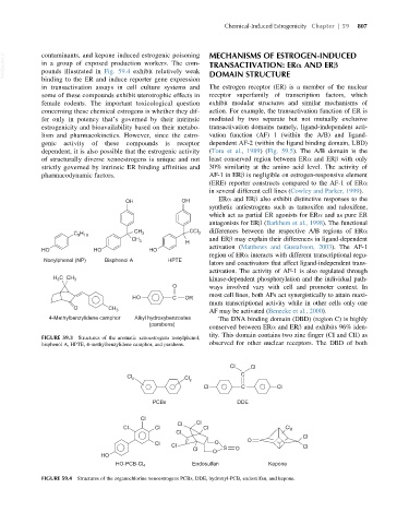
             pounds illustrated in Fig. 59.4 exhibit relatively weak
             binding to the ER and induce reporter gene expression  DOMAIN STRUCTURE
             in transactivation assays in cell culture systems and  The estrogen receptor (ER) is a member of the nuclear
             some of these compounds exhibit uterotrophic effects in  receptor superfamily of transcription factors, which
             female rodents. The important toxicological question  exhibit modular structures and similar mechanisms of
             concerning these chemical estrogens is whether they dif-  action. For example, the transactivation function of ER is
             fer only in potency that’s governed by their intrinsic  mediated by two separate but not mutually exclusive
             estrogenicity and bioavailability based on their metabo-  transactivation domains namely, ligand-independent acti-
             lism and pharmacokinetics. However, since the estro-  vation function (AF) 1 (within the A/B) and ligand-
             genic  activity  of  these  compounds  is  receptor  dependent AF-2 (within the ligand binding domain, LBD)
             dependent, it is also possible that the estrogenic activity  (Tora et al., 1989)(Fig. 59.5). The A/B domain is the
             of structurally diverse xenoestrogens is unique and not  least conserved region between ERα and ERβ with only
             strictly governed by intrinsic ER binding affinities and  30% similarity at the amino acid level. The activity of
             pharmacodynamic factors.                           AF-1 in ERβ is negligible on estrogen-responsive element
                                                                (ERE) reporter constructs compared to the AF-1 of ERα
                                                                in several different cell lines (Cowley and Parker, 1999).
                                       OH              OH         ERα and ERβ also exhibit distinctive responses to the
                                                                synthetic antiestrogens such as tamoxifen and raloxifene,
                                                                which act as partial ER agonists for ERα and as pure ER
                                                                antagonists for ERβ (Barkhem et al., 1998). The functional
                       C H               CH 3             CCl 3  differences between the respective A/B regions of ERα
                        9 19
                                        CH 3             H      and ERβ may explain their differences in ligand-dependent
                                                                activation (Matthews and Gustafsson, 2003). The AF-1
             HO              HO               HO
                                                                region of ERα interacts with different transcriptional regu-
              Nonylphenol (NP)  Bisphenol A        HPTE
                                                                lators and coactivators that affect ligand-independent trans-
                                                                activation. The activity of AF-1 is also regulated through
                 H C CH 3                                       kinase-dependent phosphorylation and the individual path-
                  3
                                                     O          ways involved vary with cell and promoter context. In
                                                                most cell lines, both AFs act synergistically to attain maxi-
                                         HO          C  OR
                                                                mum transcriptional activity while in other cells only one
                       O         CH 3                           AF may be activated (Benecke et al., 2000).
                4-Methylbenzylidene camphor  Alkyl hydroxybenzoates  The DNA binding domain (DBD) (region C) is highly
                                              (parabens)
                                                                conserved between ERα and ERβ and exhibits 96% iden-
                                                                tity. This domain contains two zinc finger (CI and CII) as
             FIGURE 59.3 Structures of the aromatic xenoestrogens nonylphenol,
             bisphenol A, HPTE, 4-methylbenzylidene camphor, and parabens.  observed for other nuclear receptors. The DBD of both


                                                                      Cl    Cl
                                       Cl x             Cl y             C
                                                              Cl         C          Cl

                                               PCBs                     DDE

                                           Cl
                                                      Cl    Cl
                                      Cl       Cl             Cl                      Cl 8
                                                      Cl
                                                                                            Cl
                                               Cl                O         O
                                                    Cl                                      Cl
                                                           Cl    O  S  O
                               HO
                                    HO-PCB-Cl 4            Endosulfan            Kepone
             FIGURE 59.4 Structures of the organochlorine xenoestrogens PCBs, DDE, hydroxyl-PCB, endosulfan, and kepone.

