
Page 1218 - Small Animal Internal Medicine, 6th Edition
P. 1218
1190 PART X Joint Disorders
VetBooks.ir
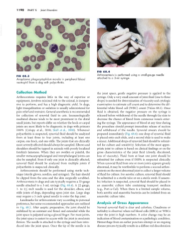
FIG 68.4
Arthrocentesis is performed using a small-gauge needle
FIG 68.3
Anaplasma phagocytophilum morula in peripheral blood attached to a 3-mL syringe.
neutrophil from a dog with polyarthritis.
Collection Method the joint space, gentle negative pressure is applied to the
Arthrocentesis requires little in the way of expertise or syringe. Only a very small amount of joint fluid (one to three
equipment, involves minimal risk to the animal, is inexpen- drops) is needed for determination of viscosity and cytologic
sive to perform, and has a high diagnostic yield. In dogs, examination to estimate cell count and to determine the dif-
light tranquilization or sedation is usually administered for ferential white blood cell (WBC) count (Video 68.1). Once
pain relief and restraint. General anesthesia is recommended fluid is obtained, the negative pressure on the syringe is
for collection of synovial fluid in cats. Immunologically released before withdrawal of the needle through the skin to
mediated disease tends to be most prominent in the distal decrease the chance of blood from cutaneous vessels enter-
small joints, but reports differ on whether the hock or carpal ing the syringe. The appearance of blood at any time during
joints are most likely to be diagnostic in dogs with primary the procedure should prompt immediate release of suction
IMPA (Colopy et al., 2010; Stull et al., 2008). Whenever and withdrawal of the needle. Synovial smears should be
polyarthritis is suspected, synovial fluid should be analyzed prepared immediately (Fig. 68.6); one drop of synovial fluid
from at least three to four joints, including at least one is placed onto each slide, and a second slide is used to make
carpus, one hock, and one stifle. The joints that are clinically a smear. Additional drops of synovial fluid should be submit-
most severely affected should always be sampled. Elbows and ted for culture and sensitivity. Selection of the most appro-
shoulders should be tapped in animals with poorly localized priate joint to culture is based on clinical findings or on the
forelimb lameness. When they are swollen or painful, the gross characteristics of the joint fluid (cloudy, discolored,
smaller metacarpophalangeal and interphalangeal joints can loss of viscosity). Fluid from at least one joint should be
also be sampled. Even if only one joint is clinically affected, submitted for culture even if IMPA is suspected clinically.
synovial fluid should be analyzed from multiple joints if When synovial fluid from one or more joints appears grossly
polyarthritis is suspected clinically. abnormal, it may be worthwhile to perform a second arthro-
Arthrocentesis should be performed using sterile tech- centesis on the most abnormal joint to collect a larger volume
nique (sterile gloves, needles, and syringes). The hair should of fluid for culture. For aerobic culture, synovial fluid should
be clipped from the area and the skin surgically scrubbed. be submitted in a sterile tube or on a sterile swab. If anaero-
Arthrocentesis in dogs and cats typically requires a 25-gauge bic infection is suspected, synovial fluid should be placed in
needle attached to a 3-mL syringe (Fig. 68.4). A 22-gauge, an anaerobic culture tube containing transport medium
1- to 1 2 -inch needle is used for the shoulder, elbow, and (e.g., Port-a-Cul). When there is a limited sample volume,
1
stifle joints of dogs, depending upon joint size. Large dogs both aerobic and anaerobic bacteria can be isolated from an
may require a 3-inch spinal needle to enter the hip joint. anaerobic culture tube.
Landmarks for arthrocentesis vary according to personal
preference, but some recommended approaches are outlined Analysis of Gross Appearance
in Fig. 68.5. After aseptic preparation, the joint should be Normal synovial fluid is clear and colorless. Cloudiness or
stabilized by an assistant and flexed and extended while the turbidity is observed when red blood cells (RBCs) or WBCs
joint space is palpated using a gloved finger. For most joints, enter the joint in high numbers. A color change may be an
the joint space is easiest to access with the joint in moderate indication of blood contamination or a pathologic condition.
flexion. The needle is attached to the syringe and then intro- Hemorrhage from an earlier puncture attempt or an ongoing
duced into the joint space. Once the tip of the needle is in disease process typically results in a diffuse red discoloration