
Page 1221 - Small Animal Internal Medicine, 6th Edition
P. 1221
CHAPTER 68 Clinical Manifestations of and Diagnostic Tests for Joint Disorders 1193
VetBooks.ir
A B
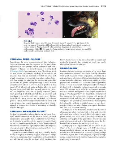
FIG 68.9
Synovial fluid from an adult German Shepherd dog with polyarthritis. (A) Some of the
cells are lupus erythematosus (LE) cells containing phagocytized, opsonized, amorphous
nuclear material. Finding these LE cells supports a diagnosis of systemic lupus
erythematosus (SLE). (B) This dog also has proteinuria, tongue ulcers caused by vasculitis,
and a positive antinuclear antibody test.
SYNOVIAL FLUID CULTURE biopsy. Needle biopsy of the synovial membrane is quick and
Bacteria are the most common cause of joint infection. minimally traumatic, but samples are small and easily
Septic arthritis can often be diagnosed on the basis of the obtained only from the stifle joint.
appearance of toxic changes within neutrophils and iden-
tification of bacteria on stained smears of synovial fluid RADIOGRAPHY
(Clements, 2005). Some organisms (e.g., Mycoplasma spp.) Radiography is an important component of the initial diag-
do not induce characteristic cytologic abnormalities, so nostic evaluation when only one joint is clinically affected or
any joint fluid with an increased nucleated cell count and when joint palpation reveals crepitation, instability, or a
a high percentage of neutrophils warrants a culture. Syno- restricted range of motion. Physical examination findings
vial fluid should be submitted for aerobic and anaerobic should be used to determine which joints should be radio-
culture and for specific Mycoplasma spp. culture. Because graphed, and two views (i.e., lateral and anterior/posterior)
direct bacterial culture of synovial fluid is positive in less should be made of each joint. Radiographic abnormalities of
than half of all cases of septic arthritis, failure to grow the joints and periarticular region are expected in animals
bacteria in synovial fluid does not rule out septic arthri- with DJD, chronic septic arthritis, and erosive immune-
tis. The diagnostic yield can be greatly improved (50%- mediated arthritis. Radiographs of the carpus are abnormal
100% positive) if infected synovial fluid is collected and in up to 75% of dogs with erosive polyarthritis at the time of
inoculated into broth-enrichment media (e.g., thiogly- first evaluation for polyarthritis. In dogs with presumed
colate blood culture bottles) and incubated for 24 hours nonerosive IMPA and dogs with infectious polyarthritis,
prior to culture. Microbiologic culture of blood, urine, and radiographs are generally not recommended if the response
synovial membrane biopsy specimens should also be con- to treatment is rapid and complete, because the only abnor-
sidered to improve the chance of recovering a clinically malities observed are mild effusion, joint capsule distention,
relevant pathogen. and associated soft tissue swelling.
Radiographs of the thorax and abdomen and abdominal
SYNOVIAL MEMBRANE BIOPSY ultrasound are often recommended in dogs and cats with
Performing synovial membrane biopsy can support a diag- polyarthritis to evaluate for underlying infectious or neo-
nosis already suspected on the basis of history, physical plastic disease that could lead to reactive polyarthritis. In
examination, radiographic studies, and synovial fluid analy- addition, radiographs of the spine should be performed in
sis. It may also be used to collect a sample for microbiologic dogs with concurrent neck or back pain to screen for dis-
culture in cases of suspected septic arthritis. Examination of kospondylitis as a cause for reactive polyarthritis.
the synovial membrane is especially valuable in the diagnosis Radiography is an important tool but has limited utility.
of neoplasia and in differentiating infectious arthritis from Many of the bony changes seen with DJD and erosive
immune-mediated disorders. immune-mediated disease are not apparent for weeks to
Synovial membrane biopsies may be obtained by needle months after the onset of signs. Although positive findings
biopsy or surgical arthrotomy. Surgical excision of a wedge contribute a great deal to the diagnosis, negative findings
of synovial membrane allows visualization of the entire joint should be interpreted with caution. Sequential radiographic
and selection of a specific site from which to obtain the studies may be warranted.