
Page 1219 - Small Animal Internal Medicine, 6th Edition
P. 1219
CHAPTER 68 Clinical Manifestations of and Diagnostic Tests for Joint Disorders 1191
VetBooks.ir
E
A
F
B
G
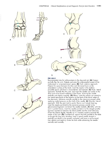
FIG 68.5
Recommended sites for arthrocentesis in the dog and cat. (A) Carpus:
partially flex the joint. Palpate and enter the anteromedial aspect of the
radiocarpal or carpometacarpal space. (B) Hock: anterior approach.
Palpate the space between the tibia and tibiotarsal bone on the
anterolateral surface of the hock; insert the needle in the shallow
C palpable space until bone is encountered, and aspirate. (C) Hock: lateral
approach. Partially flex the joint, and insert the needle just caudal to the
distal end of the lateral malleolus of the fibula, directing the needle
medially and slightly cranially. (D) Elbow: insert the needle just medial to
the lateral epicondyle of the humerus at the level of the dorsal edge of
the olecranon. Advance cranially parallel to the olecranon process while
applying medial pressure on the shaft of the needle. (E) Shoulder: lateral
approach. With the joint held in partial flexion as if weight bearing,
insert the needle just distal to the acromion process cranial to the
glenohumeral ligament and direct the needle medially. (F) Stifle: with the
joint in partial flexion, insert the needle just lateral to the straight patellar
ligament equidistant between the distal patella and the tibial tuberosity.
Direct the needle slightly medially as it is inserted caudally towards the
center of the joint. (G) Coxofemoral: support the limb parallel to the table
as though the dog were standing. Insert a spinal needle straight in
D medially just dorsal to the greater trochanter until bone is encountered,
then abduct and medially rotate the limb while advancing the needle
ventrally and caudally.