
Page 1220 - Small Animal Internal Medicine, 6th Edition
P. 1220
1192 PART X Joint Disorders
VetBooks.ir
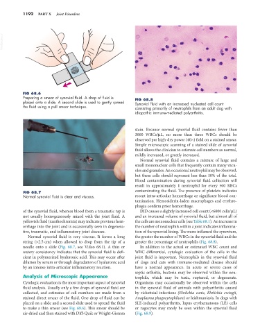
FIG 68.6
Preparing a smear of synovial fluid. A drop of fluid is FIG 68.8
placed onto a slide. A second slide is used to gently spread Synovial fluid with an increased nucleated cell count
the fluid using a pull smear technique. consisting primarily of neutrophils from an adult dog with
idiopathic immune-mediated polyarthritis.
stain. Because normal synovial fluid contains fewer than
3000 WBCs/µL, no more than three WBCs should be
observed per high-dry power (40×) field on a stained smear.
Simple microscopic scanning of a stained slide of synovial
fluid allows the clinician to estimate cell numbers as normal,
mildly increased, or greatly increased.
Normal synovial fluid contains a mixture of large and
small mononuclear cells that frequently contain many vacu-
oles and granules. An occasional neutrophil may be observed,
but these cells should represent less than 10% of the total.
Blood contamination during synovial fluid collection will
result in approximately 1 neutrophil for every 500 RBCs
FIG 68.7 contaminating the fluid. The presence of platelets indicates
Normal synovial fluid is clear and viscous. recent intra-articular hemorrhage or significant blood con-
tamination. Hemosiderin-laden macrophages and erythro-
phagia confirm prior hemorrhage.
of the synovial fluid, whereas blood from a traumatic tap is DJD causes a slightly increased cell count (<6000 cells/µL)
not usually homogeneously mixed with the joint fluid. A and an increased volume of synovial fluid, but almost all of
yellowish fluid (xanthochromia) may indicate previous hem- the cells are mononuclear cells (see Table 68.1). An increase in
orrhage into the joint and is occasionally seen in degenera- the number of neutrophils within a joint indicates inflamma-
tive, traumatic, and inflammatory joint diseases. tion of the synovial lining. The more inflamed the synovium,
Normal synovial fluid is very viscous. It forms a long the greater the number of WBCs in the synovial fluid and the
string (>2.5 cm) when allowed to drop from the tip of a greater the percentage of neutrophils (Fig. 68.8).
needle onto a slide (Fig. 68.7, see Video 68.1). A thin or In addition to the actual or estimated WBC count and
watery consistency indicates that the synovial fluid is defi- WBC differential, cytologic evaluation of the cells in the
cient in polymerized hyaluronic acid. This may occur after joint fluid is important. Neutrophils in the synovial fluid
dilution by serum or through degradation of hyaluronic acid of dogs and cats with immune-mediated disease should
by an intense intra-articular inflammatory reaction. have a normal appearance. In acute or severe cases of
septic arthritis, bacteria may be observed within the neu-
Analysis of Microscopic Appearance trophils, which may be toxic, ruptured, or degenerate.
Cytologic evaluation is the most important aspect of synovial Organisms may occasionally be observed within the cells
fluid analysis. Usually only a few drops of synovial fluid are in the synovial fluid of animals with polyarthritis caused
collected, and estimates of cell numbers are made from a by rickettsial infections (Ehrlichia canis, Ehrlichia ewingii,
stained direct smear of the fluid. One drop of fluid can be Anaplasma phagocytophilum) or leishmaniasis. In dogs with
placed on a slide and a second slide used to spread the fluid SLE-induced polyarthritis, lupus erythematosus (LE) cells
to make a thin smear (see Fig. 68.6). This smear should be or ragocytes may rarely be seen within the synovial fluid
air-dried and then stained with Diff-Quik or Wright-Giemsa (Fig. 68.9).