
Page 694 - Small Animal Internal Medicine, 6th Edition
P. 694
666 PART V Urinary Tract Disorders
Red Blood Cells cells in the urine sediment does not have localizing value.
Occasional RBCs are considered normal in the urine sedi- Caudate cells are transitional cells with tapered ends thought
VetBooks.ir ment. Normal values are: voided sample, 0 to 8/hpf; catheter- to originate from the renal pelvis. Occasional transitional
cells are normal, and increased numbers may be present if
ized sample, 0 to 5/hpf; and, cystocentesis sample, 0 to 3/hpf.
Excessive numbers of RBCs in urine is termed hematuria
(Fig. 39.2) and may be microscopic or macroscopic. The there is infection, irritation, or neoplasia of the urinary tract.
causes of hematuria are summarized in Box 39.1.
White Blood Cells
Occasional white blood cells are considered normal in the
urine sediment. Normal values are as follows: voided sample,
0 to 8/hpf; catheterized sample, 0 to 5/hpf; and cystocentesis
sample, 0 to 3/hpf. An increased number of white blood cells
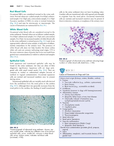
in the urine sediment is termed pyuria (Fig. 39.3) and, in an
appropriately collected urine sample, is indicative of inflam-
mation somewhere in the urinary tract. The presence of
white blood cells does not help localize the lesion unless
white cell casts are present, indicating renal origin. UTI is
the most common cause of pyuria, but it also can result from
genital tract contamination in voided or catheterized samples
(Box 39.2).
FIG 39.3
Epithelial Cells Photomicrograph of abnormal urine sediment showing large
Both squamous and transitional epithelial cells may be numbers of white blood cells (Sedi-Stain, ×100).
found in the urine sediment, but they are often of little
diagnostic significance. Squamous cells are large poly-
gonal cells with small round nuclei (Fig. 39.4). They are BOX 39.1
common in voided or catheterized samples because of
urethral or vaginal contamination. Occasional squamous Causes of Hematuria in Dogs and Cats
cells are normal and increased numbers may be present Urinary tract origin (kidneys, ureters, bladder, urethra)
during estrus. • Trauma
Transitional epithelial cells are variably sized cells derived • Traumatic collection (e.g., catheter, cystocentesis)
from the urothelium from the renal pelvis to the urethra • Renal biopsy
(Fig. 39.5). Although their size generally increases from the • Blunt trauma (e.g., automobile accident)
renal pelvis to the urethra, the finding of small transitional • Urolithiasis
• Neoplasia (common in dogs with hemangiosarcoma)
• Inflammatory disease
• Urinary tract infection
• Idiopathic feline lower urinary tract disease
• Chemically induced inflammation (e.g.,
cyclophosphamide-induced cystitis)
• Parasites
• Dioctophyma renale
• Capillaria plica
• Coagulopathy
• Warfarin intoxication
• Disseminated intravascular coagulation
• Thrombocytopenia
• Renal infarction
• Renal pelvic hematoma
• Vascular malformation
• Renal telangiectasia (Welsh Corgi)
• Idiopathic renal hematuria
FIG 39.2 Genital tract contamination (prostate gland, prepuce,
Photomicrograph of abnormal urine sediment. Arrows, top vagina)
and middle center, indicate two different sizes of transitional • Estrus
epithelial cells; arrow, middle left, indicates a white blood • Inflammatory, neoplastic, and traumatic lesions of the
cell; arrow, bottom center, indicates red blood cells genital tract
(unstained, ×100).