
Page 698 - Small Animal Internal Medicine, 6th Edition
P. 698
670 PART V Urinary Tract Disorders
VetBooks.ir
A B
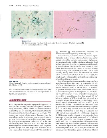
FIG 39.15
(A) Calcium oxalate monohydrate (unstained) and calcium oxalate dihydrate crystals (B)
in urine sediment (Sedi-Stain, ×400).
spp., Klebsiella spp., and Pseudomonas aeruginosa are
observed less commonly in dogs and rarely in cats.
Results obtained by bacterial culture of urine are depen-
dent on the method of urine collection. Voided urine has the
greatest potential for bacterial contamination. Catheteriza-
tion may inoculate the bladder with bacteria from the distal
urethra, but urine collected by cystocentesis should be sterile
in normal animals. Quantitative bacterial culture of urine
allows determination of the number of bacterial colonies
(colony-forming units [cfu]) that grow from 1 mL of urine
(cfu/mL). Ideally urine should be submitted for culture
within 30 minutes of collection. If this is not possible, the
sample may be refrigerated for up to 24 hours without sig-
nificant loss of bacterial growth.
FIG 39.16 Bacterial culture of midstream voided urine samples from
Photomicrograph showing cystine crystals in urine sediment normal dogs and cats often results in growth of < 10 to ≥
3
(Sedi-Stain, ×400). 10 cfu/mL. Therefore culture of voided urine is not recom-
5
mended for the evaluation of patients for UTI. If, however,
may occur in diabetes mellitus or nephrotic syndrome. They no growth is obtained from a voided urine sample, UTI can
5
also may be observed in cats because of the degeneration of be excluded as a diagnosis. Bacterial growth of ≥ 10 cfu/mL
lipid-laden tubular cells. may result from culture of urine obtained from catheteriza-
tion in 20% of normal female dogs. Thus using ≥ 10 cfu/
5
mL as an indicator of UTI in female dogs will result in a
MICROBIOLOGY substantial number of false-positive results. Also, the proce-
dure of urethral catheterization itself may cause UTI in 20%
Clinical signs and urinalysis findings provide supportive evi- of normal female dogs. Consequently, the collection of urine
dence, but microbiology is required to diagnose UTI conclu- by cystocentesis is recommended for establishing a diagnosis
sively. The kidneys, ureters, bladder, and proximal urethra of of UTI in female dogs. Isolation of bacteria from urine col-
normal dogs and cats are sterile, whereas a resident bacterial lected by catheterization of male dogs is uncommon, and
4
flora populates the distal urethra, prepuce, and vagina. UTI ≥ 10 cfu/mL is recommended for establishing a diagnosis
occurs when bacteria colonize areas of the urinary tract that of UTI in urine samples collected by catheterization from
normally are sterile. Aerobic gram-negative bacteria account male dogs. In male and female cats, growth of ≥10 cfu/mL
3
for most UTIs in dogs and cats, and the remainder are caused in samples collected by catheterization is considered com-
by gram-positive organisms. Escherichia coli is the most patible with UTI. Urine samples obtained by cystocentesis
common organism implicated in UTIs of dogs and cats. from normal dogs and cats should yield no growth because
Other organisms isolated include Proteus spp., coagulase- this procedure bypasses the normal bacterial flora of the
positive staphylococci, and streptococci. Pasturella multo- urethra and genital tract. Consequently, results obtained by
cida occasionally is isolated from cats with UTIs. Enterobacter cystocentesis are the standard against which results obtained