
Page 695 - Small Animal Internal Medicine, 6th Edition
P. 695
CHAPTER 39 Diagnostic Tests for the Urinary System 667
BOX 39.2
VetBooks.ir Causes of Pyuria in Dogs and Cats
Urinary tract origin (kidneys, ureters, bladder, urethra)
• Infectious
• Urinary tract infection (e.g., pyelonephritis, cystitis,
urethritis)
• Noninfectious
• Urolithiasis
• Neoplasia
• Trauma
• Chemically induced (e.g., cyclophosphamide)
Genital tract contamination (e.g., prostate gland,
prepuce, vagina)
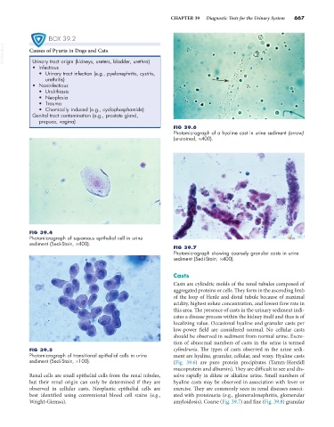
FIG 39.6
Photomicrograph of a hyaline cast in urine sediment (arrow)
(unstained, ×400).
FIG 39.4
Photomicrograph of squamous epithelial cell in urine
sediment (Sedi-Stain, ×400).
FIG 39.7
Photomicrograph showing coarsely granular casts in urine
sediment (Sedi-Stain, ×400).
Casts
Casts are cylindric molds of the renal tubules composed of
aggregated proteins or cells. They form in the ascending limb
of the loop of Henle and distal tubule because of maximal
acidity, highest solute concentration, and lowest flow rate in
this area. The presence of casts in the urinary sediment indi-
cates a disease process within the kidney itself and thus is of
localizing value. Occasional hyaline and granular casts per
low-power field are considered normal. No cellular casts
should be observed in sediment from normal urine. Excre-
tion of abnormal numbers of casts in the urine is termed
FIG 39.5 cylindruria. The types of casts observed in the urine sedi-
Photomicrograph of transitional epithelial cells in urine ment are hyaline, granular, cellular, and waxy. Hyaline casts
sediment (Sedi-Stain, ×100). (Fig. 39.6) are pure protein precipitates (Tamm-Horsfall
mucoprotein and albumin). They are difficult to see and dis-
Renal cells are small epithelial cells from the renal tubules, solve rapidly in dilute or alkaline urine. Small numbers of
but their renal origin can only be determined if they are hyaline casts may be observed in association with fever or
observed in cellular casts. Neoplastic epithelial cells are exercise. They are commonly seen in renal diseases associ-
best identified using conventional blood cell stains (e.g., ated with proteinuria (e.g., glomerulonephritis, glomerular
Wright-Giemsa). amyloidosis). Coarse (Fig. 39.7) and fine (Fig. 39.8) granular