
Page 776 - Small Animal Internal Medicine, 6th Edition
P. 776
748 PART VI Endocrine Disorders
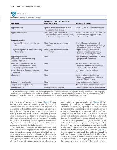
TABLE 46.4
VetBooks.ir Disorders Causing Endocrine Alopecia
ABNORMALITIES
DISORDER COMMON CLINICOPATHOLOGIC DIAGNOSTIC TESTS
Hypothyroidism Lipemia, hypercholesterolemia, mild Serum T 4, free T 4, TSH concentrations
nonregenerative anemia
Hyperadrenocorticism Stress leukogram, increased ALP, Urine cortisol/creatinine ratio, low-dose
hypercholesterolemia, hyposthenuria, dexamethasone suppression test,
proteinuria, urinary tract infection abdominal US
Hyperestrogenism
Functional Sertoli cell tumor in male None (bone marrow depression Physical findings, abdominal US,
dog uncommon) cytologic or histopathologic findings,
plasma estrogen concentration
Hyperestrogenism in intact female dog None (bone marrow depression Vaginal cytology, abdominal US,
(follicular cyst) uncommon) plasma estrogen concentration,
response to ovariohysterectomy
Hyperprogesteronism None Physical findings, abdominal US, serum
Luteal cyst in intact female dog progesterone concentrati
Adrenocortical tumor
Increased adrenocortical steroid None Measure adrenocortical steroid
hormone intermediates Occult hormone intermediates before and
(atypical) hyperadrenocorticism after ACTH administration
Growth hormone deficiency pituitary None Signalment, physical findings
dwarfism
Alopecia X* None Measure adrenocortical steroid
hormone intermediates before and
after ACTH administration
Feline endocrine alopecia None Response to progestin therapy
Telogen defluxion (effluvium) None History of recent pregnancy or diestrus
Diabetes mellitus Hyperglycemia, glycosuria Blood and urine glucose measurement
ACTH, Adrenocorticotropic hormone; ALP, alkaline phosphatase; T 4, tetraiodothyronine; TSH, thyroid-stimulating hormone; US, ultrasonography.
*Alopecia X encompasses previously named syndromes such as growth hormone–responsive dermatosis, castration-responsive dermatosis,
estrogen-responsive dermatosis, biopsy-responsive dermatosis, and congenital adrenal hyperplasia–like syndrome.
to the presence of hyperestrogenism (see Chapter 54), and tumors mimic hyperadrenocorticism (see Chapter 50). Doc-
documenting an increased plasma estrogen (i.e., estradiol) umenting increased serum progesterone concentration
concentration provides further evidence for the presence of establishes the diagnosis, especially in a male or female
a functional Sertoli cell tumor in the dog and hyperestrogen- spayed cat or dog. Serum progesterone is normally increased
ism in the bitch (assuming that the bitch is not in proestrus in an intact female dog or cat in diestrus. A history of recent
or early estrus). Abdominal ultrasound may identify ovarian cycling behavior and examination of the ovaries and adrenal
cysts or neoplasia in the bitch with hyperestrogenism, and glands with abdominal ultrasound will help differentiate
abdominal and testicular ultrasound may identify testicular diestrus, functional luteal cysts, and adrenal neoplasia.
neoplasia in the male dog. Hyperestrogenism and endocrine Alopecia X is a syndrome in dogs characterized by hair
alopecia should resolve after surgical removal of the ovarian cycle arrest, endocrine alopecia, and hyperpigmentation.
cyst, ovarian tumor, or testicular tumor. Alopecia X has been identified in many breeds, most notably
An abnormal increase in serum progesterone may result Nordic Breeds, Poodles, and “plush-coated dogs” such as the
from adrenocortical neoplasia (more common in cats than Pomeranian, Chow, Samoyed, and Keeshond (Fig. 46.4).
dogs) or functional ovarian luteal cysts in the bitch and may Alopecia occurs in young adult dogs and occurs equally in
serve as a component of an imbalance in adrenocortical males and females, intact or neutered. Systemic illness is not
steroid hormone intermediates. Functional luteal cysts may a feature of Alopecia X. Routine blood and urine test results
cause prolonged anestrus or failure to cycle in the bitch. are typically normal. Skin biopsy specimens from affected
Clinical features of progesterone-secreting adrenocortical dogs show the typical changes of endocrine alopecia (see