
Page 777 - Small Animal Internal Medicine, 6th Edition
P. 777
CHAPTER 46 Disorders of the Hypothalamus and Pituitary Gland 749
Melatonin is perhaps the least innocuous nonspecific
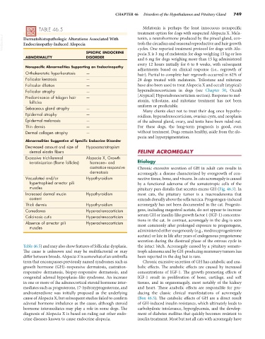
TABLE 46.5 treatment option for dogs with suspected Alopecia X. Mela-
VetBooks.ir Dermatohistopathologic Alterations Associated With tonin, a neurohormone produced by the pineal gland, con-
trols the circadian and seasonal reproductive and hair growth
Endocrinopathy-Induced Alopecia
SPECIFIC ENDOCRINE cycles. One reported treatment protocol for dogs with Alo-
pecia X is 3 mg of melatonin for dogs weighing 15 kg or less
ABNORMALITY DISORDER and 6 mg for dogs weighing more than 15 kg administered
every 12 hours initially for 6 to 8 weeks, with subsequent
Nonspecific Abnormalities Supporting an Endocrinopathy adjustments based on clinical response (i.e., regrowth of
Orthokeratotic hyperkeratosis — hair). Partial to complete hair regrowth occurred in 62% of
Follicular keratosis — 29 dogs treated with melatonin. Trilostane and mitotane
Follicular dilation — have also been used to treat Alopecia X and occult (atypical)
Follicular atrophy — hyperadrenocorticism in dogs (see Chapter 50, Occult
Predominance of telogen hair — [Atypical] Hyperadrenocorticism section). Response to mel-
follicles atonin, trilostane, and mitotane treatment has not been
Sebaceous gland atrophy — uniform or predictable.
Many clients elect not to treat their dog once hypothy-
Epidermal atrophy — roidism, hyperadrenocorticism, ovarian cysts, and neoplasia
Epidermal melanosis — of the adrenal gland, ovary, and testis have been ruled out.
Thin dermis — For these dogs, the long-term prognosis is good, even
Dermal collagen atrophy — without treatment. Dogs remain healthy, aside from the alo-
pecia and hyperpigmentation.
Abnormalities Suggestive of Specific Endocrine Disorder
Decreased amount and size of Hyposomatotropism
dermal elastin fibers FELINE ACROMEGALY
Excessive trichilemmal Alopecia X, Growth
keratinization (flame follicles) hormone– and Etiology
castration-responsive Chronic excessive secretion of GH in adult cats results in
dermatosis acromegaly, a disease characterized by overgrowth of con-
Vacuolated and/or Hypothyroidism nective tissue, bone, and viscera. In cats acromegaly is caused
hypertrophied arrector pili by a functional adenoma of the somatotropic cells of the
muscles pituitary pars distalis that secretes excess GH (Fig. 46.5). In
Increased dermal mucin Hypothyroidism most cats, the pituitary tumor is a macroadenoma that
content extends dorsally above the sella turcica. Progestogen-induced
Thick dermis Hypothyroidism acromegaly has not been documented in the cat. Progesto-
Comedones Hyperadrenocorticism gens, including megestrol acetate, do not appear to increase
Calcinosis cutis Hyperadrenocorticism serum GH or insulin-like growth factor 1 (IGF-1) concentra-
Absence of arrector pili Hyperadrenocorticism tions in the cat. In contrast, acromegaly in the dog is seen
muscles most commonly after prolonged exposure to progestogens,
administered either exogenously (e.g., medroxyprogesterone
acetate) or late in life after years of endogenous progesterone
secretion during the diestrual phase of the estrous cycle in
Table 46.5) and may also show features of follicular dysplasia. the intact bitch. Acromegaly caused by a pituitary somato-
The cause is unknown and may be multifactorial or may troph adenoma and by GH-producing mammary tumors has
differ between breeds. Alopecia X is somewhat of an umbrella been reported in the dog but is rare.
term that encompasses previously named syndromes such as Chronic excessive secretion of GH has catabolic and ana-
growth hormone (GH)–responsive dermatosis, castration- bolic effects. The anabolic effects are caused by increased
responsive dermatosis, biopsy-responsive dermatosis, and concentrations of IGF-1. The growth-promoting effects of
congenital adrenal hyperplasia–like syndrome. An increase IGF-1 result in proliferation of bone, cartilage, and soft
in one or more of the adrenocortical steroid hormone inter- tissues, and in organomegaly, most notably of the kidney
mediates such as progesterone, 17-hydroxyprogesterone, and and heart. These anabolic effects are responsible for pro-
androstenedione was initially proposed as the underlying ducing the classic clinical manifestations of acromegaly
cause of Alopecia X, but subsequent studies failed to confirm (Box 46.3). The catabolic effects of GH are a direct result
adrenal hormone imbalance as the cause, although steroid of GH-induced insulin resistance, which ultimately leads to
hormone intermediates may play a role in some dogs. The carbohydrate intolerance, hyperglycemia, and the develop-
diagnosis of Alopecia X is based on ruling out other endo- ment of diabetes mellitus that quickly becomes resistant to
crine diseases known to cause endocrine alopecia. insulin treatment. Most but not all cats with acromegaly have