
Page 782 - Small Animal Internal Medicine, 6th Edition
P. 782
754 PART VI Endocrine Disorders
pituitary stem cell after differentiation of the corticotropic BOX 46.4
cells that produce ACTH. Pituitary dwarfism resulting from
VetBooks.ir a mutant GH or insensitivity to GH owing to lack of or a Clinical Signs Associated With Pituitary Dwarfism
defect in GH receptors (e.g., Laron-type dwarfism in human
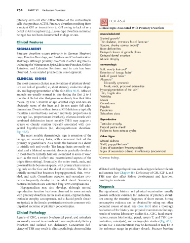
Musculoskeletal
beings) has not been documented in dogs or cats.
Stunted growth*
Clinical Features Thin skeleton, immature facial features*
Square, chunky contour (adult)*
SIGNALMENT Bone deformities
Pituitary dwarfism occurs primarily in German Shepherd Delayed closure of growth plates
Delayed dental eruption
dogs, Karelian Bear dogs, and Saarloos and Czechoslovakian Muscle atrophy
Wolfdogs, although pituitary dwarfism in other dog breeds,
including the Weimaraner, Spitz, Miniature Pinscher, Golden Dermatologic
Retreiver, and Labrador Retriever, and in cats has been Soft, woolly haircoat*
observed. A sex-related predilection is not apparent. Retention of lanugo hairs*
Lack of guard hairs*
CLINICAL SIGNS Alopecia*
The most common clinical manifestations of pituitary dwarf- Bilaterally symmetric
ism are lack of growth (i.e., short stature), endocrine alope- Trunk, neck, proximal extremities
cia, and hyperpigmentation of the skin (Box 46.4). Affected Hyperpigmentation of the skin*
animals are usually normal in size during the first 2 to 3 Thin, fragile skin
Wrinkles
months of life but after that grow more slowly than their litter Scales
mates. By 4 to 5 months of age, affected dogs and cats are Comedones
obviously runts of the litter and do not attain full adult Papules
dimensions. Dwarfs with an isolated GH deficiency typically Pyoderma
maintain a normal body contour and body proportions as Seborrhea sicca
they age (i.e., proportionate dwarfism), whereas dwarfs with
combined deficiencies (most notably TSH) may acquire a Reproductive
square or chunky contour typically associated with con- Testicular atrophy
genital hypothyroidism (i.e., disproportionate dwarfism; Flaccid penile sheath
Fig. 46.8). Failure to have estrous cycles
The most notable dermatologic sign is retention of the Other Signs
lanugo, or secondary hairs, with concurrent lack of the Mental dullness
primary or guard hairs. As a result, the haircoat in a dwarf Shrill, puppy-like bark*
is initially soft and woolly. The lanugo hairs are easily epi- Signs of secondary hypothyroidism
lated, and a bilateral symmetric alopecia gradually develops Signs of secondary adrenal insufficiency (uncommon)
in most dwarfs. Initially, hair loss is confined to areas of wear,
such as the neck (collar) and posterolateral aspects of the *Common findings.
thighs (from sitting). Eventually, the entire trunk, neck, and
proximal limbs become alopecic, with primary hairs remain-
ing only on the face and the distal extremities. The skin is affiliated with hypothyroidism, such as hypercholesterolemia
initially normal but becomes hyperpigmented, thin, wrin- and anemia (see Chapter 48). Deficiency of GH, IGF-1, and
kled, and scaly. Comedones, papules, and secondary pyo- TSH may also affect kidney development and function,
derma frequently develop in the adult dwarf. Secondary resulting in azotemia.
bacterial infections are common long-term complications.
Hypogonadism may also develop, although normal Diagnosis
reproductive function has been observed in some animals The signalment, history, and physical examination usually
with pituitary dwarfism. In the male animal, cryptorchidism, provide sufficient evidence for inclusion of pituitary dwarf-
testicular atrophy, azoospermia, and a flaccid penile sheath ism among the tentative diagnoses of short stature. Strong
are typical; in the female, persistent anestrus is common with presumptive evidence can be obtained by ruling out other
impaired secretion of pituitary gonadotropins. potential causes of small size (Box 46.5) after a thorough
evaluation of the history and physical examination findings,
Clinical Pathology results of routine laboratory studies (i.e., CBC, fecal exami-
Results of CBC, a serum biochemical panel, and urinalysis nations, serum biochemical panel, serum T 4 and TSH con-
are usually normal in animals with uncomplicated pituitary centration, urinalysis), and radiographic studies (Fig. 46.9).
dwarfism and isolated GH deficiency. Concurrent defi- Serum IGF-1 concentrations may be decreased or may be in
ciency of TSH may result in clinicopathologic abnormalities the reference range in pituitary dwarfs. Because baseline