
Page 802 - Small Animal Internal Medicine, 6th Edition
P. 802
774 PART VI Endocrine Disorders
VetBooks.ir
A B
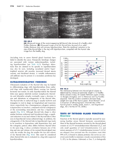
FIG 48.5
(A) Ultrasound image of the normal-appearing left thyroid lobe (arrows) of a healthy adult
Golden Retriever. (B) Ultrasound image of the left thyroid lobe (arrows) of an adult
Golden Retriever dog with primary hypothyroidism. Note the significant reduction in the
size of the thyroid lobe in the dog with hypothyroidism, compared with the thyroid lobe
image from the healthy dog.
(including tests to assess thyroid gland function) have 2,000
failed to identify the cause. Nonspecific histologic changes 1,800
are associated with various endocrinopathies, includ- 1,600
ing hypothyroidism (see Table 46.5); histologic altera- 1,400
tions that are claimed to be specific to hypothyroidism 1,200
may also be seen, including vacuolated and/or hyper- Thyroid volume (mm 3 ) 1,000
trophied arrector pili muscles, increased dermal mucin 800
content, and thickened dermis. A variable inflammatory 600
cell infiltrate may be present if a secondary pyoderma has 400
developed. 200
0
ULTRASONOGRAPHIC FINDINGS 0 10 20 30 40 50
Ultrasound evaluation of the thyroid lobe may be helpful Body weight (kg)
in differentiating dogs with hypothyroidism from euthy-
roid dogs with nonthyroidal illness causing low thyroid FIG 48.6
hormone test results. In early hypothyroidism the thyroid The relationship between total thyroid gland volume as
lobes may appear relatively normal. Lymphocytic thyroid- determined by ultrasound and body weight in 12 healthy
Akitas (closed circles), 36 Golden Retrievers (open circles),
itis and idiopathic atrophy eventually cause a decrease in 12 Beagles (triangles), and 12 Miniature and Toy Poodles
size and alterations in echogenicity of the thyroid lobe. (squares). Note the positive correlation between body
The thyroid lobe in euthyroid dogs is usually fusiform and weight and size of the thyroid gland. (From Brömel C et al:
triangular to oval in shape on longitudinal and transverse Comparison of ultrasonographic characteristics of the
views, respectively; has a homogeneous echogenic pattern; thyroid gland in healthy small-, medium-, and large-breed
is hyperechoic to isoechoic, compared with the echogenicity dogs, Am J Vet Res 67:70, 2006.)
of the surrounding musculature; and has a hyperechoic
capsule (Fig. 48.5). Although thyroid lobe shape is often
similar between euthyroid and hypothyroid dogs, a signifi- TESTS OF THYROID GLAND FUNCTION
cant reduction in size and volume of the thyroid lobe is often Overview
seen in hypothyroid versus euthyroid dogs. In addition, the Function of the thyroid gland is typically assessed by mea-
echogenicity of the thyroid lobe in hypothyroid dogs tends suring baseline serum thyroid hormone concentrations.
to be isoechoic to hypoechoic with hyperechoic foci, and Most of the thyroid hormone secreted by the thyroid gland
the echogenic pattern often differs between thyroid lobes consists of 3,5,3′5′-tetraiodothyronine (thyroxine [T 4 ]), and
in the same dog. A direct correlation between size of the only small quantities of 3,5,3′-triiodothyronine (T 3 ) and
dog and size and volume of the normal thyroid gland may minor amounts of 3,3′,5′-triiodothyronine (reverse T 3 [rT 3 ])
exist; the smaller the dog, the smaller the size and volume are released. Once secreted into the circulation, more than
of the thyroid lobe (Fig. 48.6). This must be considered 99% of T 4 is bound to plasma proteins; this serves as a res-
when thyroid lobe size is evaluated in a dog with suspected ervoir and a buffer to maintain a steady concentration of free
hypothyroidism. T 4 (fT 4 ) in the plasma. Unbound, or free, T 4 is biologically