
Page 803 - Small Animal Internal Medicine, 6th Edition
P. 803
CHAPTER 48 Disorders of the Thyroid Gland 775
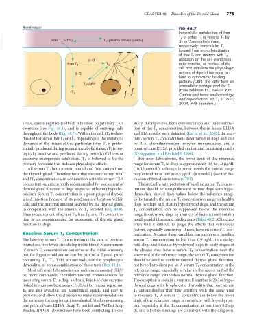
Blood vessel FIG 48.7
Intracellular metabolism of free
VetBooks.ir Free T (<1%) T –plasma protein (>99%) 5′- or 5-monodeiodinase,
T 4 to either T 3 or reverse T 3 by
4
4
respectively. Intracellular T 3
formed from monodeiodination
of free T 4 can interact with T 3
5′–D receptors on the cell membrane,
Free T 4 T 3 mitochondria, or nucleus of the
cell and stimulate the physiologic
5–D actions of thyroid hormone or
bind to cytoplasmic binding
proteins (CBP). The latter form an
T –CBP intracellular storage pool for T 3 .
3
(From Feldman EC, Nelson RW:
rT 3
Canine and feline endocrinology
and reproduction, ed 3, St Louis,
2004, WB Saunders.)
active, exerts negative feedback inhibition on pituitary TSH study, discrepancies, both overestimation and underestima-
secretion (see Fig. 48.1), and is capable of entering cells tion of the T 4 concentration, between the in house ELISA
throughout the body (Fig. 48.7). Within the cell, fT 4 is deio- and RIA results were detected (Lurye et al., 2002). In con-
dinated to form either T 3 or rT 3 , depending on the metabolic trast, serum T 4 concentrations determined in dogs and cats
demands of the tissues at that particular time. T 3 is prefer- by RIA, chemiluminescent enzyme immunoassay, and a
entially produced during normal metabolic states; rT 3 is bio- point-of-care ELISA provided similar and consistent results
logically inactive and produced during periods of illness or (Kemppainen and Birchfield, 2006).
excessive endogenous catabolism. T 3 is believed to be the For most laboratories, the lower limit of the reference
primary hormone that induces physiologic effects. range for serum T 4 in dogs is approximately 0.8 to 1.0 µg/dL
All serum T 4 , both protein bound and free, comes from (10-13 nmol/L), although in some breeds the normal range
the thyroid gland. Therefore tests that measure serum total may extend to as low as 0.5 µg/dL (6 nmol/L) (see the dis-
and fT 4 concentrations, in conjunction with the serum TSH cussion of breed variations, p. 781).
concentration, are currently recommended for assessment of Theoretically, interpretation of baseline serum T 4 concen-
thyroid gland function in dogs suspected of having hypothy- tration should be straightforward in that dogs with hypo-
roidism. Serum T 3 concentration is a poor gauge of thyroid thyroidism should have values below the reference range.
gland function because of its predominant location within Unfortunately, the serum T 4 concentration range in healthy
cells and the minimal amount secreted by the thyroid gland dogs overlaps with that in hypothyroid dogs, and the serum
in comparison with the amount of T 4 secreted (Fig. 48.8). T 4 concentration can be suppressed below the reference
Thus measurement of serum T 3 , free T 3 , and rT 3 concentra- range in euthyroid dogs by a variety of factors, most notably
tion is not recommended for assessment of thyroid gland nonthyroidal illness and medications (Table 48.2). Clinicians
function in dogs. often find it difficult to judge the effects that extraneous
factors, especially concurrent illness, have on serum T 4 con-
Baseline Serum T 4 Concentration centration. Because these variables can suppress a baseline
The baseline serum T 4 concentration is the sum of protein- serum T 4 concentration to less than 0.5 µg/dL in a euthy-
bound and free levels circulating in the blood. Measurement roid dog, and because hypothyroid dogs in early stages of
of serum T 4 concentration can serve as the initial screening the disease may have a serum T 4 concentration near the
test for hypothyroidism or can be part of a thyroid panel lower end of the reference range, the serum T 4 concentration
containing T 4 , fT 4 , TSH, an antibody test for lymphocytic should be used to confirm normal thyroid gland function,
thyroiditis, or some combination of these tests (Box 48.4). not hypothyroidism per se. A serum T 4 concentration in the
Most reference laboratories use radioimmunoassay (RIA) reference range, especially a value in the upper half of the
or, more commonly, chemiluminescent immunoassays for reference range, establishes normal thyroid gland function.
measuring serum T 4 in dogs and cats. Point-of-care enzyme- The exception is seen in a very small number (<2%) of hypo-
linked immunosorbent assays (ELISAs) for measuring serum thyroid dogs with lymphocytic thyroiditis that have serum
T 4 are also available; are economical, quick, and easy to T 4 autoantibodies that may interfere with the assay used
perform; and allow the clinician to make recommendations to measure T 4 . A serum T 4 concentration below the lower
the same day the dog (or cat) is evaluated. Studies evaluating limit of the reference range is consistent with hypothyroid-
one point-of-care ELISA (Snap T 4 test kit and VetTest Snap ism, especially if the T 4 concentration is less than 0.5 µg/
Reader, IDDEX laboratories) have been conflicting. In one dL and all other findings are consistent with the diagnosis.