
Page 828 - Small Animal Internal Medicine, 6th Edition
P. 828
800 PART VI Endocrine Disorders
Approximately 5% of cats require a second I treatment. nodes and lungs are common. Metastasis to other locations
131
Factors such as dose of radioactive iodine administered, such as the liver, kidney, bone, and brain is also possible.
VetBooks.ir extent of abnormal thyroid tissue, iodine excretion rate, and Most dogs with thyroid tumor are euthyroid or hypothyroid;
less than 15% of dogs have functional thyroid tumors that
thyroid pathology may explain treatment failure. Most wor-
risome is the possibility of thyroid carcinoma. Cats with
thyroid carcinoma require higher doses of radioactive iodine secrete excess thyroid hormone, causing hyperthyroidism.
Clinical signs of hyperthyroidism may predominate in these
than is typically administered to attain a successful outcome. dogs. Hyperthyroidism may be caused by functional thyroid
The duration of hospitalization and home care of the cat fol- adenomas and carcinomas. Adenomatous hyperplasia is the
lowing I administration varies depending on state regula- most common cause of hyperthyroidism in cats but has not
131
131
tions and the dosage of I administered. Hyperthyroidism been described in dogs.
may recur 1 year or longer after successful treatment.
Clinical Features
Prognosis Thyroid tumors occur in middle-aged to older dogs, typically
The prognosis is excellent for most cats with hyperthyroid- 10 years of age and older. No sex-related predilection has
ism, as long as concurrent disease can be managed and been noted. Although any breed can be affected, Boxers,
131
thyroid carcinoma is not the cause. Surgery and I therapy Beagles, Golden Retrievers, and Siberian Huskies may be at
have the potential for cure, although hyperthyroidism may increased risk.
recur months to years (or not at all) after thyroidectomy or Dogs with nonfunctional thyroid tumors are usually
131 I treatment. Hyperthyroid cats with adenomatous hyper- brought to veterinarians because the owner has seen or
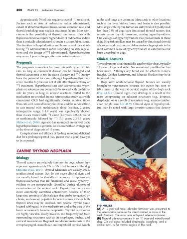
plasia or adenoma can potentially be treated with methima- felt a mass in the ventral cervical region of the dog’s neck
zole for years, as long as adverse reactions related to the (Fig. 48.22). Clinical signs may develop as a result of the
medication are avoided. In one retrospective study, cats with mass compressing on adjacent structures (e.g., dyspnea,
concurrent CKD had significantly shorter survival times dysphagia) or as a result of metastasis (e.g., exercise intoler-
than cats with normal kidney function, and the survival time ance, weight loss; Box 48.9). Clinical signs of hypothyroid-
in cats treated with methimazole alone (median, 2 years; ism may be noted with large invasive tumors that destroy
interquartile range, 1-3.9 years) was significantly shorter
131
than in cats treated with I alone (4.0 years; 3.0-4.8 years)
or methimazole followed by 131 I (5.3 years; 2.2-6.5 years;
Milner et al., 2006). Age also has an impact on survival data;
hyperthyroidism is a geriatric cat disease with an average age
at the time of diagnosis of 13 years.
Complications and efficacy of feeding an iodine-deficient
diet for a prolonged period (i.e., greater than a year) have yet
to be reported.
CANINE THYROID NEOPLASIA
A
Etiology
Thyroid tumors are relatively common in dogs, where they
represent approximately 1% to 3% of all tumors in the dog
(Broome et al., 2014). Thyroid adenomas are usually small,
nonfunctional masses that do not cause clinical signs and
are usually found incidentally at necropsy. Exceptions are
thyroid adenomas that are functional and cause hyperthy-
roidism or are unexpectedly identified during ultrasound
examination of the ventral neck. Thyroid carcinomas are
more commonly identified antemortem because of their
large size, presence of clinical signs that can be recognized by
clients, and ease of palpation by veterinarians. One or both B
thyroid lobes may be involved, and ectopic thyroid tissue
located sublingual, in the mediastinum and at the base of the FIG 48.22
heart occasionally become neoplastic. Thyroid carcinomas (A) A 13-year-old male Labrador Retriever was presented to
the veterinarian because the client noticed a mass in the
are highly vascular, locally invasive, and frequently infiltrate neck (arrows). The mass was a thyroid adenocarcinoma.
surrounding structures such as the esophagus, trachea, and (B) Thyroid adenocarcinoma in an 11-year-old mixed-breed
cervical musculature. Regional and distant metastasis to the dog. Clinical signs included dysphagia, coughing, and a
retropharyngeal, mandibular, and superficial cervical lymph visible mass in the ventral region of the neck.