
Page 829 - Small Animal Internal Medicine, 6th Edition
P. 829
CHAPTER 48 Disorders of the Thyroid Gland 801
BOX 48.9
VetBooks.ir Clinical Signs Caused by Thyroid Neoplasia in Dogs
Nonfunctional
Swelling or mass in neck
Dyspnea
Cough
Lethargy
Dysphagia
Regurgitation
Anorexia
Weight loss
Horner syndrome
Change in bark
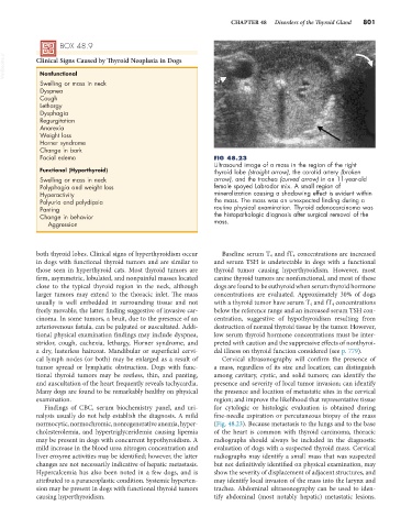
Facial edema FIG 48.23
Ultrasound image of a mass in the region of the right
Functional (Hyperthyroid) thyroid lobe (straight arrow), the carotid artery (broken
Swelling or mass in neck arrow), and the trachea (curved arrow) in an 11-year-old
Polyphagia and weight loss female spayed Labrador mix. A small region of
Hyperactivity mineralization causing a shadowing effect is evident within
Polyuria and polydipsia the mass. The mass was an unexpected finding during a
Panting routine physical examination. Thyroid adenocarcinoma was
Change in behavior the histopathologic diagnosis after surgical removal of the
Aggression mass.
both thyroid lobes. Clinical signs of hyperthyroidism occur Baseline serum T 4 and fT 4 concentrations are increased
in dogs with functional thyroid tumors and are similar to and serum TSH is undetectable in dogs with a functional
those seen in hyperthyroid cats. Most thyroid tumors are thyroid tumor causing hyperthyroidism. However, most
firm, asymmetric, lobulated, and nonpainful masses located canine thyroid tumors are nonfunctional, and most of these
close to the typical thyroid region in the neck, although dogs are found to be euthyroid when serum thyroid hormone
larger tumors may extend to the thoracic inlet. The mass concentrations are evaluated. Approximately 30% of dogs
usually is well embedded in surrounding tissue and not with a thyroid tumor have serum T 4 and fT 4 concentrations
freely movable; the latter finding suggestive of invasive car- below the reference range and an increased serum TSH con-
cinoma. In some tumors, a bruit, due to the presence of an centration, suggestive of hypothyroidism resulting from
arteriovenous fistula, can be palpated or auscultated. Addi- destruction of normal thyroid tissue by the tumor. However,
tional physical examination findings may include dyspnea, low serum thyroid hormone concentrations must be inter-
stridor, cough, cachexia, lethargy, Horner syndrome, and preted with caution and the suppressive effects of nonthyroi-
a dry, lusterless haircoat. Mandibular or superficial cervi- dal illness on thyroid function considered (see p. 779).
cal lymph nodes (or both) may be enlarged as a result of Cervical ultrasonography will confirm the presence of
tumor spread or lymphatic obstruction. Dogs with func- a mass, regardless of its size and location; can distinguish
tional thyroid tumors may be restless, thin, and panting, among cavitary, cystic, and solid tumors; can identify the
and auscultation of the heart frequently reveals tachycardia. presence and severity of local tumor invasion; can identify
Many dogs are found to be remarkably healthy on physical the presence and location of metastatic sites in the cervical
examination. region; and improve the likelihood that representative tissue
Findings of CBC, serum biochemistry panel, and uri- for cytologic or histologic evaluation is obtained during
nalysis usually do not help establish the diagnosis. A mild fine-needle aspiration or percutaneous biopsy of the mass
normocytic, normochromic, nonregenerative anemia, hyper- (Fig. 48.23). Because metastasis to the lungs and to the base
cholesterolemia, and hypertriglyceridemia causing lipemia of the heart is common with thyroid carcinoma, thoracic
may be present in dogs with concurrent hypothyroidism. A radiographs should always be included in the diagnostic
mild increase in the blood urea nitrogen concentration and evaluation of dogs with a suspected thyroid mass. Cervical
liver enzyme activities may be identified; however, the latter radiographs may identify a small mass that was suspected
changes are not necessarily indicative of hepatic metastasis. but not definitively identified on physical examination, may
Hypercalcemia has also been noted in a few dogs, and is show the severity of displacement of adjacent structures, and
attributed to a paraneoplastic condition. Systemic hyperten- may identify local invasion of the mass into the larynx and
sion may be present in dogs with functional thyroid tumors trachea. Abdominal ultrasonography can be used to iden-
causing hyperthyroidism. tify abdominal (most notably hepatic) metastatic lesions.