
Page 830 - Small Animal Internal Medicine, 6th Edition
P. 830
802 PART VI Endocrine Disorders
Computed tomographic (CT) and magnetic resonance canine thyroid tumors are highly vascular, and it is common
imaging (MRI) can define the extent of tumor invasion into for hemorrhage to occur after biopsy. Fine-needle aspiration
VetBooks.ir surrounding structures, identify distant metastasis to the using a 21- or 23-gauge needle and cytologic examination of
the mass are recommended initially to confirm that the mass
lymph nodes and lung, and identify ectopic thyroid tissue in
the mediastinum (Fig. 48.24)—information that is valuable
blood is common, and differentiation between adenoma and
if surgery or radiation therapy is being considered. Both CT is of thyroid origin. Contamination of the aspirate with
and MRI are superior to ultrasound for establishing extent carcinoma is difficult. Large-bore needle biopsy, surgical
of invasion of thyroid tumors. exploration, or ultrasound-guided biopsy is often required
Thyroid scans using sodium pertechnetate can be used to to confirm the diagnosis. Ultrasonography identifies solid
confirm that a cervical mass is thyroid in origin; to assess the areas of the mass to biopsy and large blood vessels to be
degree of regional tissue invasion; and to identify unusual avoided. This procedure is preferred if the findings yielded
areas of uptake in the head, neck, and thorax suggestive of by needle aspiration are inconclusive.
metastatic sites. Most thyroid carcinomas demonstrate het-
erogeneous uptake of pertechnetate, irregular gland shape, Treatment
and evidence of regional tissue invasion. Tumors with a more Treatment options for thyroid tumor in dogs include surgery,
homogenous diffuse uptake of pertechnetate are more likely chemotherapy, radiation therapy, radioactive iodine treat-
to be functional tumors that cause hyperthyroidism. If the ment, and antithyroid drugs. In many cases more than one
malignancy, especially a distant site of metastasis, does not treatment option (e.g., surgery and chemotherapy) is used
trap iodine effectively, the scintigraphic study will fail to either concurrently or sequentially.
identify the site. Failure to identify distant metastatic sites The therapeutic approach is based, in part, on the size and
with scintigraphy does not mean that distant metastasis does invasiveness of the tumor and the presence of regional and
not exist. The amount of radionuclide uptake by the thyroid distant metastasis. Approximately 30% to 40% of dogs with
tumor is not a reliable indicator of its functional status (i.e., thyroid tumors have detectable distant metastasis at the time
euthyroid, hypothyroid, or hyperthyroid) or of the benign of diagnosis. Tumor size appears to be predictive of meta-
3
versus malignant nature of the tumor. Thoracic radiographs static behavior, with thyroid tumor volume less than 21 cm
are more sensitive than a thyroid scan in identifying pulmo- having a significantly lower risk of metastasis and thyroid
nary metastasis. tumor volume approaching 100 cm having essentially a
3
100% probability of metastasis. The functional status of the
Diagnosis thyroid tumor does not dramatically alter the treatment
Definitive diagnosis can be obtained either by doing a fine approach. All thyroid tumors in dogs should be considered
needle aspirate for cytology or a biopsy. Unfortunately, malignant until proven otherwise, regardless of size (Fig.
48.25). Treatment is warranted even for large, locally invasive
tumors. Many dogs with large invasive tumors appear more
0
1
2
3
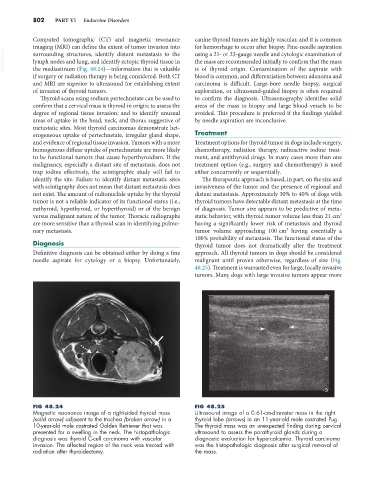
FIG 48.24 FIG 48.25
Magnetic resonance image of a right-sided thyroid mass Ultrasound image of a 0.61-cm-diameter mass in the right
(solid arrow) adjacent to the trachea (broken arrow) in a thyroid lobe (arrows) in an 11-year-old male castrated Pug.
10-year-old male castrated Golden Retriever that was The thyroid mass was an unexpected finding during cervical
presented for a swelling in the neck. The histopathologic ultrasound to assess the parathyroid glands during a
diagnosis was thyroid C-cell carcinoma with vascular diagnostic evaluation for hypercalcemia. Thyroid carcinoma
invasion. The affected region of the neck was treated with was the histopathologic diagnosis after surgical removal of
radiation after thyroidectomy. the mass.