Page 255 - Withrow and MacEwen's Small Animal Clinical Oncology, 6th Edition
P. 255
234   PART III    Therapeutic Modalities for the Cancer Patient


         T-cell tolerance rather than activate T cells. 91–93  The DCs present   immunosuppressive cytokines. 106  A  few key immunosuppres-
         in the tumor tend to be immature and dysfunctional. Studies of   sive cytokines produced by tumor cells are IL-10, VEGF, and
                                                                      106,107
                                                               TGF-β.
                                                                           These cytokines act to suppress antitumor T cell
         DCs in numerous human cancers demonstrate minimal activa-
  VetBooks.ir  tion, decreased ability to stimulate in an alloreactive fashion, and   responses and inhibit DC function. IL-10 promotes Treg pro-
                                                       A simi-
                                                                                    and, in an autocrine and/or paracrine
                                               90,91,94–99
         decreased expression of costimulatory molecules.
                                                                                 108
                                                               duction and function
         lar study done in dogs with canine transmissible venereal tumors   fashion, may potentially affect tumor cell proliferation and sur-
         (CTVTs) showed that the tumor environment caused downregula-  vival. 109  In human cancer patients, increased levels of serum
         tion of DC surface markers of activation and MHC, and decreased   IL-10 are observed in patients with either pancreatic carcinoma
         endocytic capabilities and decreased allogenic mixed lymphocyte   or non-Hodgkin lymphoma (NHL). 110,111  In addition, elevated
         reaction (MLR) responses.  Possible mechanisms causing the DC   levels of IL-10 in diffuse large B-cell lymphoma of humans cor-
                              56
         dysfunction include the overexpression of the protein S100A9, 100    relate with a poor prognosis. 112  TGF-β acts similarly to IL-10 in
         accumulation of triglycerides in the DCs which leads to decreased   that it is a potent immunosuppressive cytokine that can potenti-
         capacity to present antigen, 101  and downregulation of toll-like   ate Treg proliferation and function. 74,113–115  It can also enhance
         receptor (TLR)-9 expression. 102  Moreover, factors such as IL-10   tumor progression; carcinomas can produce excess  TGF-β,
         and vascular endothelial growth factor (VEGF) can negatively   which in turn increases the epithelial-to-mesenchymal transi-
         affect DC function and maturation. 103,104  Lastly, some DCs in   tion, tumor invasion, and metastasis and inhibits tumor-specific
         the tumor are considered to be regulatory based on low expres-  CD8+ T  cells. 115  Moreover, tumor-produced tumor necrosis
         sion of surface markers MHC II, CD86, and CD11c with high   factor-alpha (TNF-α) leads to promotion of tumor cell survival
         expression of costimulatory molecules CD80, CD40, CD106,   via induction of antiapoptotic proteins. 116  Finally, TNF-α has
         and CD11b. These cells secrete regulatory factors such as IL-10   been shown to promote tumor angiogenesis and metastasis and
         and nitric oxide (NO) and inhibit proliferation of naïve CD4+ T   hamper cytotoxic T-cell and macrophage responses. 117
         cells to antigen presented by mature, functional DCs. 105  Overall,   One study in veterinary medicine examined a lymph node
         the microenvironment of the tumor leads to attraction of imma-  of a dog with metastatic melanoma. This study revealed an
         ture and regulatory DCs that, due to their decreased activation   overexpression of IL-10 and TGF-β concurrent with a lack of
         and function, can potently inhibit the development of antitumor    expression of IL-2, IL-4, or interferon-gamma (IFN-γ) cytokines
         T cell responses even when copious amounts of antigen are present.   typically associated with antitumor immunity, thus demonstrat-
                                                               ing that tumor immunosuppression occurs in veterinary patients
         Production of Immunosuppressive Cytokines             as well. 118  For a review of cytokines relevant to tumor immuno-
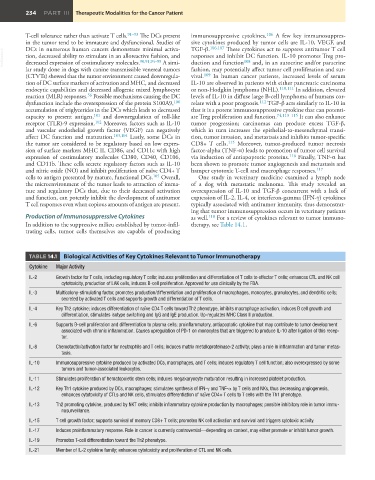
         In addition to the suppressive milieu established by tumor-infil-  therapy, see Table 14.1.
         trating cells, tumor cells themselves are capable of producing


          TABLE 14.1     Biological Activities of Key Cytokines Relevant to Tumor Immunotherapy
           Cytokine  Major Activity
           IL-2    Growth factor for T cells, including regulatory T cells; induces proliferation and differentiation of T cells to effector T cells; enhances CTL and NK cell
                     cytotoxicity, production of LAK cells, induces B-cell proliferation. Approved for use clinically by the FDA.
           IL-3    Multicolony-stimulating factor, promotes production/differentiation and proliferation of macrophages, monocytes, granulocytes, and dendritic cells;
                     secreted by activated T cells and supports growth and differentiation of T cells.
           IL-4    Key Th2 cytokine; induces differentiation of naïve CD4 T cells toward Th2 phenotype, inhibits macrophage activation, induces B cell growth and
                     differentiation, stimulates isotype switching and IgG and IgE production. Up-regulates MHC Class II production.
           IL-6    Supports B-cell proliferation and differentiation to plasma cells; proinflammatory, antiapoptotic cytokine that may contribute to tumor development
                     associated with chronic inflammation. Causes upregulation of PD-1 on monocytes that are triggered to produce IL-10 after ligation of this recep-
                     tor.
           IL-8    Chemotactic/activation factor for neutrophils and T cells; induces matrix metalloproteinase-2 activity; plays a role in inflammation and tumor metas-
                     tasis.
           IL-10   Immunosuppressive cytokine produced by activated DCs, macrophages, and T cells; induces regulatory T cell function; also overexpressed by some
                     tumors and tumor-associated leukocytes.
           IL-11   Stimulates proliferation of hematopoietic stem cells; induces megakaryocyte maturation resulting in increased platelet production.

           IL-12   Key Th1 cytokine produced by DCs, macrophages; stimulates synthesis of IFN-γ and TNF-α by T cells and NKs, thus decreasing angiogenesis,
                     enhances cytotoxicity of CTLs and NK cells, stimulates differentiation of naïve CD4+ T cells to T cells with the Th1 phenotype.
           IL-13   Th2 promoting cytokine, produced by NKT cells; inhibits inflammatory cytokine production by macrophages; possible inhibitory role in tumor immu-
                     nosurveillance.
           IL-15   T cell growth factor; supports survival of memory CD8+ T cells; promotes NK cell activation and survival and triggers cytotoxic activity.
           IL-17   Induces proinflammatory response. Role in cancer is currently controversial—depending on context, may either promote or inhibit tumor growth.
           IL-19   Promotes T-cell differentiation toward the Th2 phenotype.
           IL-21   Member of IL-2 cytokine family; enhances cytotoxicity and proliferation of CTL and NK cells.

