
Page 486 - Withrow and MacEwen's Small Animal Clinical Oncology, 6th Edition
P. 486
464 PART IV Specific Malignancies in the Small Animal Patient
Contrast radiography, although used less after advances in US, walls thicker than 1 cm are nearly four times as likely to have a
has often been used to evaluate patients with signs of primary tumor and those with focal lesions are nearly 20 times as likely
to have a tumor.
524
GI disease. US can help facilitate noninvasive localization of the
Nevertheless, possible differential diagnoses
VetBooks.ir tumor and identification of other sites of metastasis or involve- include fungal (pythiosis and histoplasmosis) masses, as these can
mimic neoplasia.
ment. It also can guide needle aspiration or needle biopsy or assist
In general, neoplasia exhibits more dramatic
528
in treatment planning. US is a more sensitive diagnostic test than thickening with loss of wall layering and greater LN enlargement,
radiographs for identifying a mass. 441,451,454,523 US is also less as well as more frequent focal lesions than nonneoplastic intesti-
time consuming than contrast radiography, and the increased use, nal disease. 528 Similar changes (thickened muscularis propria, and
availability, and operator skill for the former has diminished the ratio of muscularis to mucosa >1) can be seen in cats with intes-
need for the latter. tinal lymphoma, but do not reliably distinguish neoplasia from
US findings in dogs and cats with intestinal neoplasia most IBD. 529 In a series of 14 cats with carcinomatosis, three of which
consistently include bowel wall thickening and loss of normal were a result of small intestinal tumors (two carcinomas and one
wall layering. 456,523,524 Intestinal lymphoma in dogs more often lymphoma), the hallmark ultrasonographic finding was the pres-
results in long segments of involved bowel and either a solitary ence of masses in the double sheet portion of peritoneum that
mass or diffusely thickened bowel loops with thickening of the connects the visceral and parietal portions (100% of cats); all cats
muscularis propria in cats. 506,524,525 However, the normal appear- also had free peritoneal fluid. 530
ance of intestine does not rule out the presence of lymphoma, as
one study showed 26% of dogs diagnosed with GI lymphoma Thoracic Radiographs
did not have sonographic abnormalities. 526 Adenocarcinoma in Thoracic radiographs are critical to the complete evaluation of
cats has been described as having mixed echogenicity and was the cancer patient. For dogs with nonlymphomatous intesti-
asymmetric in three of five cats. 523 In one study, two-thirds of nal tumors, yield is low, with very few patients presenting with
dogs with intestinal adenocarcinoma had hypoechoic tumors and pulmonary metastasis. 441 This may be due to a bias in reporting
most had decreased motility. 456 These masses averaged 4 cm long because many reports detail outcome of treatment and patients
with a median wall thickness of 1.2 cm. 456,473 MCTs have an with metastatic disease may not receive treatment. In fact, many
eccentric appearance with alteration, but not loss of wall layering, case series report no evidence of metastasis on initial evaluation
commonly involving the muscularis propria. 522 Smooth muscle for solid tumors of the intestine in dogs. 441,451,452,456,458 Two of
tumors are characteristically large (median diameter 4.8 cm) and 14 cats in one series and no cats in another series had pulmo-
anechoic/hypoechoic, and a muscular layer origin may be identi- nary nodules at initial evaluation. 444,458 For cats and dogs with
fied. Leiomyomas may have a smooth contour. 454 One report of lymphoma, enlarged sternal or perihilar LNs, pleural effusion, or
metastatic mammary carcinoma to the small intestine described diffuse interstitial changes may be seen. 445,450
the appearance as multiple, hypoechoic, well-defined or margin-
ated nodules within the muscularis layer of the jejunum that did Endoscopy, Colonoscopy, and Laparoscopy
not disrupt the intestinal layering. 527 Minimally invasive methods of collecting tissues to aid in diag-
Degree of thickening, distribution of lesion(s), and symmetry nosis are increasingly used. Endoscopic findings in dogs with
are used to help differentiate neoplastic from nonneoplastic dis- intestinal lymphoma include an irregular cobblestone or patchy
ease. 528 In one study, 99% of dogs with neoplasia had a loss of wall erythematous appearance to the duodenal mucosa and poor dis-
layering and this was associated with a 50 times greater likelihood tensibility and elasticity of the duodenal wall. 453 Colonoscopy can
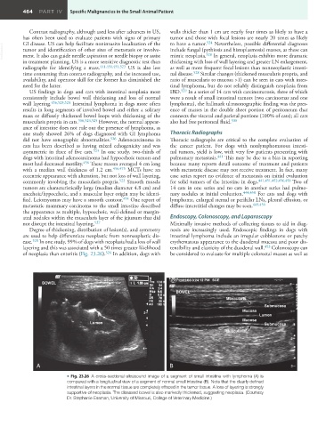
of neoplasia than enteritis (Fig. 23.26). 524 In addition, dogs with be considered to evaluate for multiple colorectal masses as well as
BOWEL
BOWEL
Muscularis
Submucosa
Mucosa
Lumen
Mucosa
Lumen Submucosa
Muscularis
A B
• Fig. 23.26 A cross-sectional ultrasound image of a segment of small intestine with lymphoma (A) is
compared with a longitudinal view of a segment of normal small intestine (B). Note that the clearly defined
intestinal layers in the normal tissue are completely effaced in the tumor tissue. A loss of layering is strongly
supportive of neoplasia. The diseased bowel is also markedly thickened, suggesting neoplasia. (Courtesy
Dr. Stephanie Essman, University of Missouri, College of Veterinary Medicine.)