
Page 487 - Withrow and MacEwen's Small Animal Clinical Oncology, 6th Edition
P. 487
CHAPTER 23 Cancer of the Gastrointestinal Tract 465
obtain biopsy samples; however, single rectal masses appear to be with chemotherapy except when intestinal perforation or the need
more common in dogs and these masses were not present beyond for a biopsy necessitates surgery (Fig. 23.28). As long as severe
Therefore proctoscopy or transanal single
extraserosal invasion and/or adhesions do not complicate the sur-
531
the colorectal border.
VetBooks.ir laparoscopic port evaluation may provide information regarding gical approach, complete excision of intestinal tumors is often
possible. For dogs and cats without evidence of local or distant
mass number and characteristics without the need for extensive
bowl preparation and surgical delay. 531 Of note, 5 of 16 dogs metastasis, long-term survival is possible, although some tumors
(31%) had different colonoscopy biopsy results compared with may later metastasize. Overall, the 1-year survival rate is approxi-
the final histopathology results with a tendency to underdiagnose mately 40% for dogs with solid small intestinal tumors. 441 For
malignancy. 531 Interobserver variation is likely to be more pro- cats with adenocarcinoma, approximately 50% will metastasize
nounced with small tissue samples and this is a limitation of these to the local LNs, 30% to the peritoneal cavity (carcinomatosis),
less invasive approaches. and 20% or less to the lungs. 434,458,462 Dogs have similar rates
of metastasis to LNs for both adenocarcinoma and leiomyosar-
Exploratory Laparotomy coma, although the liver is usually the second most frequent
When noninvasive or minimally invasive diagnostics fail to con- site. 441,458,473 Perioperative mortality can approach 30% to 50%
firm a diagnosis, an exploratory laparotomy may be indicated for as a result of sepsis, peritonitis, or owner decision for euthanasia
dogs and cats with persistent signs of GI disease. Benefits include when nonresectable tumors are present. 441,451
direct visualization of all abdominal viscera and the ability to col-
lect full-thickness biopsies of all segments of intestines and other Small Intestine
viscera. Patients with resectable solid tumors may be both diag- Intestinal resection and anastomosis is the most common surgical
nosed and treated in a single procedure with intestinal resection technique for tumors of the small intestine. Stapling techniques
and anastomosis. In a series of dogs with GI lymphoma, endo- have been shown to be equivalent to hand suturing in both the
scopic biopsies were sometimes difficult to interpret because of large and small intestine. 533,534 Canine small intestinal adenocar-
lymphoplasmacytic infiltrate, but surgical biopsies obtained by cinoma has a guarded prognosis with a mean survival time (ST) of
laparotomy confirmed the diagnosis in all cases. 450 In a study only 12 days without treatment and a mean ST of 114 days after
evaluating 367 dogs and cats undergoing GI biopsies, the risk of surgical resection, though others report median STs (MSTs) of 7
GI dehiscence was found to be very low (1% dogs, <3% cats) and 10 months. 441,456,458 Dogs with leiomyosarcoma who survive
with possible risk factors in cats being neoplasia and hypoalbu- the perioperative period have MSTs of 1.1 to almost 2 years. 451,452
menia, although these had wide confidence intervals. 532 It should One case series found the MST for 28 dogs with GIST to be
be noted that carcinomatosis should not always be seen as an indi- approximately 38 months (1 year if postoperative deaths were
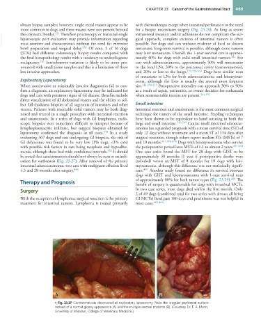
cation for euthanasia (Fig. 23.27). After removal of the primary included) versus an MST of 8 months for 10 dogs with leio-
intestinal adenocarcinoma, two cats with malignant effusion lived myosarcoma, although this difference was not statistically signifi-
4.5 and 28 months after surgery. 444 cant. 487 Another study found no difference in survival between
dogs with GIST and leiomyosarcoma with 1-year survival rates
Therapy and Prognosis of approximately 80% for both tumor types (Fig. 23.29). 488 The
benefit of surgery is questionable for dogs with intestinal MCTs.
Surgery In two case series, most dogs died within the first month. Only
2 of 49 dogs (combined total for two series with almost all being
With the exception of lymphoma, surgical resection is the primary GI MCTs) lived past 180 days and prednisone was not helpful in
treatment for intestinal tumors. Lymphoma is treated primarily most cases. 465,466
A B
• Fig. 23.27 Carcinomatosis discovered at exploratory laparotomy. Note the irregular peritoneal surface
instead of a normal glossy appearance (A) and the multiple serosal implants (B). (Courtesy Dr. F. A. Mann,
University of Missouri, College of Veterinary Medicine.)