
Page 483 - Withrow and MacEwen's Small Animal Clinical Oncology, 6th Edition
P. 483
CHAPTER 23 Cancer of the Gastrointestinal Tract 461
Pathology and Natural Behavior lymphoma is often a systemic disease; 25% of dogs and 80%
of cats will have concurrent involvement of other organs. 443,450
Epithelial, mesenchymal, neuroendocrine, and discrete/round
VetBooks.ir cell neoplasias can all be found in the intestinal tract. Although Lymphoma
most small intestinal tumors are malignant in dogs, most rec-
tal tumors are benign polyps, adenomas, or carcinomas in situ Lymphoma is the most common type of small intestinal neoplasia
(Fig. 23.24). 455,468 in cats and dogs. For feline intestinal lymphoma, subtypes include
When tumors of the GI system metastasize, sites of predilec- lymphocytic, lymphoblastic, epitheliotropic, and large granular
tion in decreasing frequency include mesenteric LNs (especially lymphocyte (LGL) types. Intestinal lymphoma in dogs occurs in
adenocarcinoma), liver (especially leiomyosarcoma), mesentery, the stomach and small intestine equally and more often in both of
omentum, spleen, kidney, bone, peritoneum (e.g., carcino- these sites than in the large intestine. For additional information
matosis), and lung. 441,458,462,469 Interestingly, metastasis from regarding canine and feline lymphoma the reader is referred to
intestinal adenocarcinoma was discovered in three dogs initially Chapter 33, Sections A and B.
presented for testicular masses. 470 One dog was presented for
multiple cutaneous masses that IHC confirmed were epithelial Adenomatous Polyps and Adenocarcinoma
in origin and a primary small intestinal adenocarcinoma with
additional visceral metastasis was diagnosed at necropsy. 471 GI Most alimentary adenocarcinoma in cats is found in the
small intestine 433,456,462 ; however, the colon and rectum are
more common sites in dogs. 472,473 For colorectal adenocarci-
nomas, the rectum is a more common site than the colon. 474
The cecum is more likely to develop leiomyosarcomas or
GISTs than adenocarcinoma. 451,473 Histologic descriptors for
carcinoma of the intestine include adeno- (forming glands),
mucinous (>50% mucin), signet ring (>50% of cells have intra-
cellular mucin), and undifferentiated or solid (no evidence of
gland formation). 472 Grossly, colorectal adenocarcinomas may
demonstrate a pedunculated (especially in the distal rectum),
cobblestone (middle rectum), or annular (middle rectum)
appearance, which may relate to behavior and prognosis (Fig.
23.25). 469,473,474
Adenomatous polyps are found in the rectum of dogs and carci-
nomas in situ are found in both the colon and rectum. Most lesions
are solitary, although multiple and diffuse lesions can be seen and
are associated with increased recurrence rates. 455 A case series of
31 dogs with colorectal carcinoma found that most were B-cell,
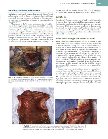
• Fig. 23.24 Cobblestone appearance to a rectal adenocarcinoma. Dogs 475
with this tumor type live an average of 12 months after surgical excision. high-grade, and caused hematochezia. Miniature dachshunds
(Courtesy Dr. Eric Pope, Ross University, College of Veterinary Medi- are overrepresented for inflammatory colorectal polyps, suggesting
cine.) a breed predisposition in multiple case series from Japan. 476,477 The
A B
• Fig. 23.25 An annular form of colonic adenocarcinoma causing a structure. The thick band of tissue (B)
creating the stricture is seen on cross-section (A). In one study, dogs with this type of tumor survived an
average of only 1.6 months. (Courtesy Dr. Eric Pope, Ross University, College of Veterinary Medicine.)