
Page 480 - Withrow and MacEwen's Small Animal Clinical Oncology, 6th Edition
P. 480
458 PART IV Specific Malignancies in the Small Animal Patient
a useful, minimally invasive technique to obtain cellular or tis-
sue samples for diagnostic purposes. 398–401 A coagulation profile
is recommended before hepatic biopsy because mild-to-moder-
VetBooks.ir ate hemorrhage is the most frequent complication, occurring in
398–401
approximately 5% of cases.
A correct diagnosis is obtained
in up to 60% of hepatic aspirates and 90% of needle-core biop-
sies. 398–401,407 The most useful cytologic features for the diagnosis
of well-differentiated HCCs include dissociation of hepatocytes,
acinar or palisading arrangement of neoplastic cells, and the pres-
ence of naked nuclei and capillaries, together with mild anisocyto-
sis, anisokaryosis, multinuclearity, and increased N:C ratios. 408,409
More invasive techniques, such as laparoscopy and open keyhole
approaches, can also be used for the biopsy and staging of cats and
dogs with suspected liver tumors. In humans, laparoscopy is rec-
ommended for local staging, as up to 20% of cases do not proceed
with open surgery because of either nodular or diffuse tumors or
unresectable disease. 410 However, for solitary and massive hepatic
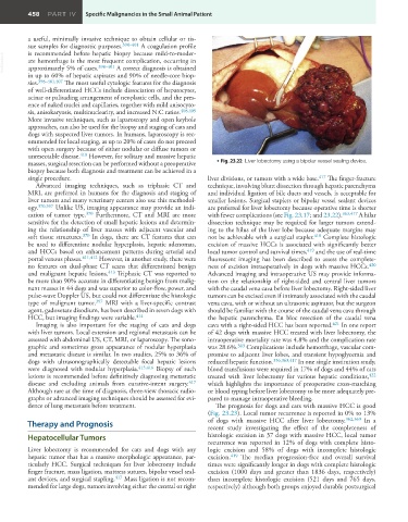
masses, surgical resection can be performed without a preoperative • Fig. 23.22 Liver lobectomy using a bipolar vessel sealing device.
biopsy because both diagnosis and treatment can be achieved in a
single procedure. liver divisions, or tumors with a wide base. 417 The finger-fracture
Advanced imaging techniques, such as triphasic CT and technique, involving blunt dissection through hepatic parenchyma
MRI, are preferred in humans for the diagnosis and staging of and individual ligation of bile ducts and vessels, is acceptable for
liver tumors and many veterinary centers also use this methodol- smaller lesions. Surgical staplers or bipolar vessel sealant devices
ogy. 370,397 Unlike US, imaging appearance may provide an indi- are preferred for liver lobectomy because operative time is shorter
cation of tumor type. 370 Furthermore, CT and MRI are more with fewer complications (see Fig. 23.17; and 23.22). 363,417 A hilar
sensitive for the detection of small hepatic lesions and determin- dissection technique may be required for larger tumors extend-
ing the relationship of liver masses with adjacent vascular and ing to the hilus of the liver lobe because adequate margins may
soft tissue structures. 370 In dogs, there are CT features that can not be achievable with a surgical stapler. 418 Complete histologic
be used to differentiate nodular hyperplasia, hepatic adenomas, excision of massive HCCs is associated with significantly better
and HCCs based on enhancement patterns during arterial and local tumor control and survival times, 419 and the use of real-time
portal venous phases. 411,412 However, in another study, there were fluorescent imaging has been described to assess the complete-
no features on dual-phase CT scans that differentiated benign ness of excision intraoperatively in dogs with massive HCCs. 420
and malignant hepatic lesions. 413 Triphasic CT was reported to Advanced imaging and intraoperative US may provide informa-
be more than 90% accurate in differentiating benign from malig- tion on the relationship of right-sided and central liver tumors
nant masses in 44 dogs and was superior to color-flow, power, and with the caudal vena cava before liver lobectomy. Right-sided liver
pulse-wave Doppler US, but could not differentiate the histologic tumors can be excised even if intimately associated with the caudal
type of malignant tumor. 397 MRI with a liver-specific contrast vena cava, with or without an ultrasonic aspirator, but the surgeon
agent, gadoxetate disodium, has been described in seven dogs with should be familiar with the course of the caudal vena cava through
HCC, but imaging findings were variable. 414 the hepatic parenchyma. En bloc resection of the caudal vena
Imaging is also important for the staging of cats and dogs cava with a right-sided HCC has been reported. 421 In one report
with liver tumors. Local extension and regional metastasis can be of 42 dogs with massive HCC treated with liver lobectomy, the
assessed with abdominal US, CT, MRI, or laparoscopy. The sono- intraoperative mortality rate was 4.8% and the complication rate
graphic and sometimes gross appearance of nodular hyperplasia was 28.6%. 363 Complications include hemorrhage, vascular com-
and metastatic disease is similar. In two studies, 25% to 36% of promise to adjacent liver lobes, and transient hypoglycemia and
dogs with ultrasonographically detectable focal hepatic lesions reduced hepatic function. 356,363,417 In one single institution study,
were diagnosed with nodular hyperplasia. 415,416 Biopsy of such blood transfusions were required in 17% of dogs and 44% of cats
lesions is recommended before definitively diagnosing metastatic treated with liver lobectomy for various hepatic conditions, 422
disease and excluding animals from curative-intent surgery. 417 which highlights the importance of preoperative cross-matching
Although rare at the time of diagnosis, three-view thoracic radio- or blood typing before liver lobectomy to be more adequately pre-
graphs or advanced imaging techniques should be assessed for evi- pared to manage intraoperative bleeding.
dence of lung metastasis before treatment. The prognosis for dogs and cats with massive HCC is good
(Fig. 23.23). Local tumor recurrence is reported in 0% to 13%
Therapy and Prognosis of dogs with massive HCC after liver lobectomy. 362,363 In a
recent study investigating the effect of the completeness of
Hepatocellular Tumors histologic excision in 37 dogs with massive HCC, local tumor
recurrence was reported in 12% of dogs with complete histo-
Liver lobectomy is recommended for cats and dogs with any logic excision and 58% of dogs with incomplete histologic
hepatic tumor that has a massive morphologic appearance, par- excision. 419 The median progression-free and overall survival
ticularly HCC. Surgical techniques for liver lobectomy include times were significantly longer in dogs with complete histologic
finger fracture, mass ligation, mattress sutures, bipolar vessel seal- excision (1000 days and greater than 1836 days, respectively)
ant devices, and surgical stapling. 417 Mass ligation is not recom- than incomplete histologic excision (521 days and 765 days,
mended for large dogs, tumors involving either the central or right respectively) although both groups enjoyed durable postsurgical