
Page 479 - Withrow and MacEwen's Small Animal Clinical Oncology, 6th Edition
P. 479
CHAPTER 23 Cancer of the Gastrointestinal Tract 457
tumors. 353–366,373 With respect to the massive form of HCC, most TABLE 23.7 Common Clinicopathologic
clinical signs are related to the mechanical mass effect of the tumor Abnormalities in Cats and Dogs with
and only rarely to any systemic effects of the tumor or hepatic
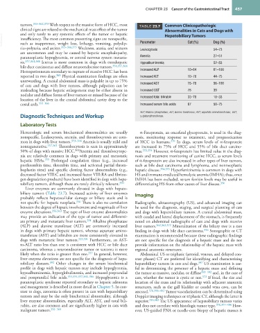
VetBooks.ir insufficiency. The most common presenting signs are nonspecific, Parameter Hepatobiliary Tumors Dog (%)
Cat (%)
such as inappetence, weight loss, lethargy, vomiting, polydip-
sia–polyuria, and ascites. 353–366,373 Weakness, ataxia, and seizures Leukocytosis 54–73
are uncommon and may be caused by hepatic encephalopathy,
paraneoplastic hypoglycemia, or central nervous system metasta- Anemia 27–51
sis. 357,361,388 Icterus is more common in dogs with extrahepatic Hypoalbuminemia 52–83
bile duct carcinomas and diffuse neuroendocrine tumors. 354,357,364
Hemoperitoneum secondary to rupture of massive HCC has been Increased ALP 10–64 61–100
reported in two dogs. 389 Physical examination findings are often Increased ALT 10–78 44–75
unrewarding. A cranial abdominal mass is palpable in up to 75%
of cats and dogs with liver tumors, although palpation can be Increased AST 15–78 56–100
misleading because hepatic enlargement may be either absent in Increased GGT 78 39
nodular and diffuse forms of liver tumors or missed because of the
location of the liver in the cranial abdominal cavity deep to the Increased total bilirubin 33–78 18–33
costal arch. 353–366 Increased serum bile acids 67 50–75
ALP, Alkaline phosphatase; ALT, alanine transferase; AST, aspartate aminotransferase; GGT,
Diagnostic Techniques and Workup γ-glutamyltransferase.
Laboratory Tests
Hematologic and serum biochemical abnormalities are usually α-Fetoprotein, an oncofetal glycoprotein, is used in the diag-
nonspecific. Leukocytosis, anemia, and thrombocytosis are com- nosis, monitoring response to treatment, and prognostication
mon in dogs with liver tumors. 353–366 Anemia is usually mild and of HCC in humans. 370 In dogs, serum levels of α-fetoprotein
nonregenerative. 357,363 Thrombocytosis is seen in approximately are increased in 75% of HCC and 55% of bile duct carcino-
50% of dogs with massive HCC. 363 Anemia and thrombocytope- mas. 393,394 However, α-fetoprotein has limited value in the diag-
nia are relatively common in dogs with primary and metastatic nosis and treatment monitoring of canine HCC, as serum levels
hepatic HSAs. 355 Prolonged coagulation times (e.g., increased of α-fetoprotein are also increased in other types of liver tumors,
prothrombin time, thrombin time, and activated partial throm- such as bile duct carcinoma and lymphoma, and nonneoplastic
boplastin time) and specific clotting factor abnormalities (e.g., hepatic disease. 394,395 Hyperferritinemia is common in dogs with
decreased factor VIII:C and increased factor VIII:RA and fibrino- HS and immune-mediated hemolytic anemia (IMHA); thus, once
gen degradation products) have been identified in dogs with hepa- IMHA has been excluded, serum ferritin levels may be useful in
tobiliary tumors, although these are rarely clinically relevant. 390 differentiating HS from other causes of liver disease. 396
Liver enzymes are commonly elevated in dogs with hepato-
biliary tumors (Table 23.7). Increased activity of liver enzymes Imaging
probably reflects hepatocellular damage or biliary stasis and is
not specific for hepatic neoplasia. 356 There is also no correlation Radiographs, ultrasonography (US), and advanced imaging can
between the degree of hepatic involvement and magnitude of liver be used for the diagnosis, staging, and surgical planning of cats
enzyme alterations. 356,363 The type of liver enzyme abnormalities and dogs with hepatobiliary tumors. A cranial abdominal mass,
may provide an indication of the type of tumor and differenti- with caudal and lateral displacement of the stomach, is frequently
ate primary and metastatic liver tumors. 391 Alkaline phosphatase noted on abdominal radiographs of cats and dogs with massive
(ALP) and alanine transferase (ALT) are commonly increased liver tumors. 362,363,372 Mineralization of the biliary tree is a rare
in dogs with primary hepatic tumors, whereas aspartate amino- finding in dogs with bile duct carcinoma. 356 Sonographic or CT
transferase (AST) and bilirubin are more consistently elevated in examination is recommended because these radiographic findings
dogs with metastatic liver tumors. 353,391 Furthermore, an AST- are not specific for the diagnosis of a hepatic mass and do not
to-ALT ratio less than one is consistent with HCC or bile duct provide information on the relationship of the hepatic mass with
carcinoma, whereas a neuroendocrine tumor or sarcoma is more regional anatomic structures.
likely when the ratio is greater than one. 357 In general, however, Abdominal US or triphasic (arterial, venous, and delayed con-
liver enzyme elevations are not specific for the diagnosis of hepa- trast phases) CT are preferred for identifying and characterizing
tobiliary diseases. 392 Other changes in the serum biochemical hepatobiliary tumors in cats and dogs. 397 US examination is use-
profile in dogs with hepatic tumors may include hypoglycemia, ful in determining the presence of a hepatic mass and defining
hypoalbuminemia, hyperglobulinemia, and increased preprandial the tumor as massive, nodular, or diffuse 398–402 and, in the case of
and postprandial bile acids. 353,354,357,361–366 Hypoglycemia is a cats, whether the tumor is cystic or not. 372 If focal, the size and
paraneoplastic syndrome reported secondary to hepatic adenoma location of the mass and its relationship with adjacent anatomic
and management is described in more detail in Chapter 5. In con- structures, such as the gall bladder or caudal vena cava, can be
trast to dogs, azotemia is often present in cats with hepatobiliary assessed. 376,398–402 Tumor vascularization can be determined using
tumors and may be the only biochemical abnormality, although Doppler imaging techniques or triphasic CT, although the latter is
liver enzyme abnormalities, especially ALT, AST, and total bili- superior. 356,402 The US appearance of hepatobiliary tumors varies
rubin, are also common and are significantly higher in cats with and does not correlate with histologic tumor type. 376,397–406 How-
malignant tumors. 358–360 ever, US-guided FNA or needle-core biopsy of hepatic masses is