
Page 475 - Withrow and MacEwen's Small Animal Clinical Oncology, 6th Edition
P. 475
CHAPTER 23 Cancer of the Gastrointestinal Tract 453
without cavitating ulceration, and are most often found at the
cardia or pylorus. 293,318–321 Paraneoplastic hypoglycemia has
been reported with leiomyoma and leiomyosarcoma, possi-
VetBooks.ir bly due to excessive release of IGF-2. 320,321 Although smooth
muscle tumors are more common in the stomach, approximately
10% to 20% of GISTs arise at this site. 299,304,322 GISTs arise
from the interstitial cells of Cajal, which normally express c-Kit
(CD117) and may also express CD34, and hence immunohis-
tochemistry (IHC) is required to differentiate leiomyosarcomas
from GISTs. 322,323 Mutations in exon 11 of the c-kit gene are
common in canine GIST and mutations in exon 9 have been
reported. 299,324–327 GISTs are rare in cats. 328
Gastric involvement of feline alimentary lymphoma is rela-
tively uncommon. 329,330 Readers should refer to Chapter 33, Sec-
tion B (Feline Lymphoma and Leukemia) for further information
regarding gastric lymphoma.
History and Clinical Signs • Fig. 23.15 Dorsal plane computed tomography image of a dog with an
ulcerated and cavitated pyloric leiomyosarcoma (white arrowhead).
Vomiting is the most common clinical sign, with or with-
out associated hematemesis, in cats and dogs with gastric
tumors. 284,292 Weight loss, anorexia, melena, diarrhea, and
abdominal pain may also be encountered. Gastric cancer
should be considered as a potential cause of septic peri-
tonitis or pneumoperitoneum. Duration of clinical signs
may vary widely, but is commonly in the order of 1 to 2
months. 292,294
Diagnostic Techniques and Workup
Routine blood tests are not expected to be diagnostic, but may
reveal anemia, hypoalbuminemia, thrombocytopenia, or throm-
bocytosis in patients with hemorrhage associated with ulcer-
ation. 284,292 Hepatocellular leakage enzymes may be increased
with liver metastasis. Abdominal radiographs may identify
changes such as a cranial abdominal mass, loss of serosal detail,
or apparent thickening of the gastric wall. Contrast radiogra-
phy may be helpful to identify delayed gastric emptying. Given
the limited detail typically observed on radiographs, this modal-
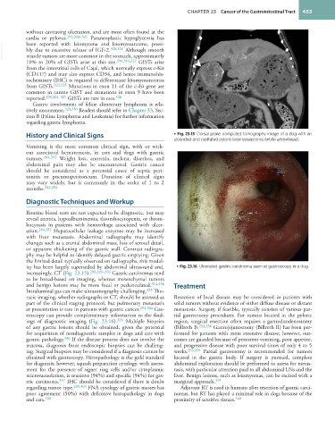
ity has been largely superseded by abdominal ultrasound and, • Fig. 23.16 Ulcerated gastric carcinoma seen at gastroscopy in a dog.
increasingly, CT (Fig. 23.15). 292,331–333 Gastric carcinomas tend
to be broad-based on imaging, whereas mesenchymal tumors
and benign lesions may be more focal or pedunculated. 314,334 Treatment
Intraluminal gas can make ultrasonography challenging. 335 Tho-
racic imaging, whether radiographs or CT, should be assessed as Resection of local disease may be considered in patients with
part of the clinical staging protocol, but pulmonary metastasis solid tumors without evidence of either diffuse disease or distant
at presentation is rare in patients with gastric cancer. 292,304 Gas- metastasis. Surgery, if feasible, typically consists of various par-
troscopy can provide complementary information to the find- tial gastrectomy procedures. For tumors located in the pyloric
ings of diagnostic imaging (Fig. 23.16). 335 Multiple biopsies region, surgical resection often requires a gastroduodenostomy
of any gastric lesions should be obtained, given the potential (Billroth I). 293,338 Gastrojejunostomy (Billroth II) has been per-
for acquisition of nondiagnostic samples in dogs and cats with formed for patients with more extensive disease; however, out-
gastric pathology. 336 If the disease process does not involve the comes are guarded because of persistent vomiting, poor appetite,
mucosa, diagnosis from endoscopic biopsies can be challeng- and progressive disease with poor survival times of only 4 to 5
ing. Surgical biopsies may be considered if a diagnosis cannot be weeks. 293,339 Partial gastrectomy is recommended for tumors
obtained with gastroscopy. Histopathology is the gold standard located in the gastric body. If surgery is pursued, complete
for diagnosis; however, squash preparation cytology, with assess- abdominal exploration should be performed to assess for metas-
ment for the presence of signet ring cells and/or cytoplasmic tasis, with particular attention paid to all abdominal LNs and the
microvacuolation, is sensitive (94%) and specific (94%) for gas- liver. Benign lesions, such as leiomyomas, can be excised with a
tric carcinoma. 337 IHC should be considered if there is doubt marginal approach. 319
regarding tumor type. 299,304 FNA cytology of gastric masses has Adjuvant RT is used in humans after resection of gastric carci-
poor agreement (50%) with definitive histopathology in dogs nomas, but RT has played a minimal role in dogs because of the
and cats. 308 proximity of sensitive tissues. 340