
Page 472 - Withrow and MacEwen's Small Animal Clinical Oncology, 6th Edition
P. 472
450 PART IV Specific Malignancies in the Small Animal Patient
VetBooks.ir
• Fig. 23.14 Lateral thoracic radiographic image of a large (8 cm long ×
6 cm wide) leiomyosarcoma in the caudal esophagus, just cranial to the
diaphragm. (From Farese JP, Bacon NJ, Ehrhart NP, et al. Oesophageal
leiomyosarcoma in dogs: surgical management and clinical outcome of
four cases. Vet Comp Oncol. 2008;6(1):31–38.)
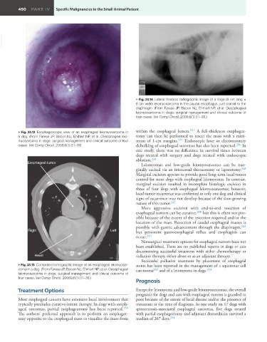
• Fig. 23.12 Esophagoscopic view of an esophageal leiomyosarcoma in within the esophageal lumen. 251 A full-thickness esophagec-
a dog. (From Farese JP, Bacon NJ, Ehrhart NP, et al. Oesophageal leio- tomy can then be performed to resect the mass with a mini-
myosarcoma in dogs: surgical management and clinical outcome of four mum of 1-cm margins. 251 Endoscopic laser or electrocautery
cases. Vet Comp Oncol. 2008;6(1):31–38) debulking of esophageal sarcomas has also been reported. 236 In
one study, there was no difference in survival times between
dogs treated with surgery and dogs treated with endoscopic
ablation. 233
Esophageal tumor
Leiomyomas and low-grade leiomyosarcomas can be mar-
ginally excised via an intercostal thoracotomy or laparotomy. 248
Marginal excision appears to provide good long-term local tumor
control for most dogs with esophageal leiomyomas. In contrast,
marginal excision resulted in incomplete histologic excision in
three of four dogs with esophageal leiomyosarcomas; however,
local tumor recurrence was confirmed in only one dog and clinical
signs of recurrence may not develop because of the slow-growing
nature of this tumor. 242
More aggressive excision with end-to-end resection of
esophageal tumors can be curative, 240 but this is often not pos-
sible because of the extent of the resection required and/or the
location of the mass. Resection of caudal esophageal masses is
possible with gastric advancement through the diaphragm, 243
Right Left but persistent gastroesophageal reflux and esophagitis can
occur. 250
Nonsurgical treatment options for esophageal tumors have not
been established. There are no published reports in dogs or cats
documenting successful treatment with either chemotherapy or
Heart radiation therapy, either alone or as an adjuvant therapy.
Successful palliative treatment by placement of esophageal
• Fig. 23.13 Computed tomographic image of an esophageal leiomyosar- stents has been reported in the management of a squamous cell
coma in a dog. (From Farese JP, Bacon NJ, Ehrhart NP, et al. Oesophageal carcinoma 252 and of a leiomyoma in dogs. 253
leiomyosarcoma in dogs: surgical management and clinical outcome of
four cases. Vet Comp Oncol. 20089;6(1):31–38.)
Prognosis
Treatment Options Except for leiomyoma and low-grade leiomyosarcomas, the overall
prognosis for dogs and cats with esophageal tumors is guarded to
Most esophageal cancers have extensive local involvement that poor because of the extent of local disease and/or the presence of
typically precludes curative-intent therapy. In dogs with esoph- metastasis at the time of diagnosis. In one study on 17 dogs with
ageal sarcomas, partial esophagectomy has been reported. 251 spirocercosis-associated esophageal sarcomas, five dogs treated
The authors’ preferred approach is to perform an esophagot- with partial esophagectomy and adjuvant doxorubicin survived a
omy opposite to the esophageal mass to visualize the mass from median of 267 days. 234