
Page 477 - Withrow and MacEwen's Small Animal Clinical Oncology, 6th Edition
P. 477
CHAPTER 23 Cancer of the Gastrointestinal Tract 455
Pathology and Natural Behavior
VetBooks.ir Hepatocellular Tumors
Hepatocellular tumors include HCC, hepatocellular adenoma
(or hepatoma), and hepatoblastoma. 356 Hepatoblastoma is a rare
tumor of primordial hepatic stem cells and has only been reported
in one dog. 369 Hepatocellular adenoma is usually an incidental
finding and rarely causes clinical signs. 354 Of the hepatocellular
tumors, hepatocellular adenoma is more common in cats and
HCC occurs more frequently in dogs. 354,357,358
HCC is the most common primary liver tumor in dogs,
accounting for up to 77% of cases, and the second most common
in cats. 354–360,367 Etiologic factors implicated in the development
of HCC in humans include infection with hepatitis virus B or
C and cirrhosis. 370 A viral etiology has also been demonstrated
in woodchucks but not in cats or dogs, and cirrhosis is rare in
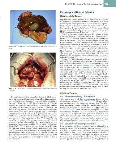
• Fig. 23.18 Nodular morphologic appearance of a bile duct carcinoma in dogs with HCC. 358–361 A link between progressive vacuolar hepa-
a cat. topathy and HCC has been proposed in Scottish terriers, with
HCC diagnosed in 34% of Scottish terriers with progressive vacu-
olar hepatopathy. 371 In one study, 20% of dogs with HCC were
diagnosed with additional tumors although most were benign and
endocrine in origin. 357
A breed and sex predisposition has not been confirmed in dogs
with HCC, but miniature schnauzers and male dogs are over-
represented in some studies. 357,361,363,372 Morphologically, 53%
to 83% of HCCs are massive (see Fig. 23.17), 16% to 25% are
nodular, and up to 19% are diffuse. 354,357 The left liver lobes,
which include the left lateral and medial lobes and papillary pro-
cess of the caudate lobe, are involved in more than two-thirds of
dogs with massive HCC, 357,361–363 but tumors are equally distrib-
uted between the left and right liver lobes in cats. 373 Metastasis
to regional LNs, peritoneum, and lungs is more common in dogs
with nodular and diffuse HCC. 354,357,361 Other metastatic sites
include the heart, kidneys, adrenal glands, pancreas, intestines,
spleen, and urinary bladder. 354,357,361 The metastatic rate varies
from 0% to 37% for dogs with massive HCCs and 93% to 100%
• Fig. 23.19 Diffuse morphologic appearance in a dog with a bile duct car- for dogs with nodular and diffuse HCCs. 354,357–363
cinoma.
Bile Duct Tumors
In people, primary liver tumors have been reclassified accord- Bile Duct Adenoma (Biliary Cystadenoma)
ing to the presence of hepatic progenitor cells and immunohisto- There are two types of bile duct tumors in cats and dogs: bile duct
chemical markers to differentiate hepatocytic and cholangiocytic adenoma and carcinoma. 354,357–360,364,365,374–378 Bile duct adeno-
lineages. 367 Liver tumors with hepatic progenitor cell charac- mas are common in cats, accounting for more than 50% of all
teristics are typically more poorly differentiated and biologi- feline hepatobiliary tumors, and are also known as biliary or hepa-
cally aggressive, resulting in a poorer prognosis. 367 Primary liver tobiliary cystadenomas because of their cystic appearance (Fig.
tumors in dogs and cats have also been reclassified according to 23.20). 358–360,374–376 Male cats may be predisposed. 374,376 Bile
these criteria. 367,368 Based on histologic findings and the degree duct adenomas usually do not cause clinical signs until they reach
of immunoreactivity to keratin 19, hepatocellular tumors were a large size and compress adjacent organs. 374–376 There is an even
divided into well-differentiated, scirrhous, and poorly differenti- distribution between single and multiple lesions. 358–360,374–376
ated tumors. 367 HCCs expressing less than 5% of keratin 19 were Malignant transformation has been reported in humans, and ana-
more likely to be derived from mature hepatocytes with minimal plastic changes have been observed in some feline adenomas. 358,374
evidence of cellular pleomorphism, infiltrative growth, and rare
metastasis, 367 and this accounted for 79% of canine HCCs and all Bile Duct Carcinoma (Cholangiocarcinoma)
feline HCCs. 367,368 In contrast, primary liver tumors with keratin Bile duct carcinoma is the most common malignant hepatobiliary
19 expression, which included poorly differentiated HCCs and tumor in cats and the second most common in dogs. 354,357–360
cholangiosarcomas, were characterized by a high grade of cellular Bile duct carcinomas account for 9% to 41% of all malignant
pleomorphism, infiltrative growth, vascular invasion, and intra- or liver tumors in dogs. 357,368,379 In humans, trematode infestation,
extrahepatic metastasis. 367,368 These tumors were probably derived cholelithiasis, and sclerosing cholangitis are known risk factors
from either hepatic progenitor cells or dedifferentiation of mature for bile duct carcinoma. 380 Trematodes may also be involved in
hepatocytes. 367 the etiology of bile duct carcinoma in cats and dogs, but they are