
Page 476 - Withrow and MacEwen's Small Animal Clinical Oncology, 6th Edition
P. 476
454 PART IV Specific Malignancies in the Small Animal Patient
The role of chemotherapy for animals with solid gastric TABLE 23.6 Morphologic Types of Canine Hepatic
tumors is unclear. Response of carcinomas to chemotherapy Tumors
has typically been poor, although multiple protocols have been
VetBooks.ir attempted. 284,292,293 Expression of HER-2 is common in canine Massive Nodular Diffuse (%)
(%)
(%)
gastric carcinoma (58%), and this may represent a therapeutic tar-
get in dogs; however, clinical data are not available. 341 Responses Hepatocellular 53–84 16–25 0–19
to imatinib have been reported in dogs with GISTs, but large-scale carcinoma
studies are lacking. 324,325
Bile duct carcinoma 37–46 0–46 17–54
Prognosis Neuroendocrine tumor 0 33 67
Gastric carcinoma typically carries a poor prognosis because of Sarcoma 36 64 0
the difficulty in achieving local tumor control and a moderate-
to-high metastatic rate. Long-term survival is possible after
partial gastrectomy, but survival times are usually less than 6 mon-
ths. 292–294,297,338,342,343 Given the challenges with controlling sys-
temic disease, few dogs with gastric carcinoma are good surgical
candidates and careful case selection is critical. Improvements in
systemic therapies may increase the number of animals consid-
ered candidates for surgery. The median survival times for dogs
with GIST, leiomyosarcoma, and undifferentiated sarcoma, pro-
vided they survive the perioperative period, are 37.4 months, 8
to 12 months, and 2.9 months, respectively. 304,318 The prognosis
is excellent after surgical resection of leiomyoma, with the major-
ity of dogs cured. 319,344 The median survival times for dogs and
cats with gastrointestinal MCTs are less than 1 month 300 and 531
days, respectively. 311
Comparative Aspects
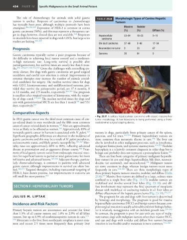
• Fig. 23.17 A solitary hepatocellular carcinoma with classic massive liver
In 2012 gastric cancer was the third most common cause of can- tumor morphology. A liver lobectomy is being performed using a thora-
cer-related death in men worldwide and the fifth most common coabdominal surgical stapling device.
cause of cancer-related death in women. 345 Men are approximately
twice as likely to be affected as women. 346 Approximately 89% of
noncardia gastric cancer in humans is associated with H. pylori. 347 tumors in dogs, particularly from primary cancer of the spleen,
Significant geographic differences are observed in the incidence of pancreas, and GI tract. 353,354 Primary hepatobiliary tumors are
gastric carcinoma and this may be a result of H. pylori prevalence, more common than metastatic disease in cats. 356 The liver can
socioeconomic status, and likely genetic susceptibility. 316,347 Mor- also be involved in other malignant processes, such as lymphoma,
tality rates are approximately 60% to 80%, reflecting advanced malignant histiocytosis, and systemic mastocytosis. 354,355 Nodular
disease at presentation and an aggressive disease course. 348 Treat- hyperplasia is a relatively common diagnosis in older dogs but is
ment of local gastric tumors varies from endoscopic mucosal resec- benign and probably does not represent a preneoplastic lesion. 356
tion for superficial lesions to partial or total gastrectomy for more There are four basic categories of primary malignant hepatobi-
infiltrative and advanced lesions. 349,350 Adjuvant therapy, particu- liary tumors in cats and dogs: hepatocellular, bile duct, neuroen-
larly chemoradiotherapy, is common in patients with advanced docrine (or carcinoid), and mesenchymal. 356 Malignant tumors
gastric cancer, although improvements in survival are often lim- are more common in dogs, whereas benign tumors occur more
ited. 340,351 Targeted therapies, including trastuzumab targeting of frequently in cats. 354–360 There are three morphologic types of
HER-2, have shown promise, but improvements in outcome are these primary hepatic tumors: massive, nodular, and diffuse (Table
still small for most patients. 352 23.6). 357 Massive liver tumors are defined as a large, solitary mass
confined to a single liver lobe (Fig. 23.17); nodular tumors are
multifocal and involve several liver lobes (Fig. 23.18); and dif-
SECTION F: HEPATOBILIARY TUMORS fuse involvement may represent the final spectrum of neoplastic
disease with multifocal or coalescing nodules in all liver lobes or
diffuse effacement of the hepatic parenchyma (Fig. 23.19). 356,357
JULIUS M. LIPTAK The prognosis for cats and dogs with liver tumors is determined
by histology and morphology. The prognosis is good for massive
Incidence and Risk Factors hepatocellular carcinomas (HCC) and benign tumors because com-
plete surgical resection is usually achievable and their biologic behav-
Primary hepatic tumors are uncommon and account for fewer ior is relatively nonaggressive with an indolent growth rate. 359–363
than 1.5% of all canine tumors and 1.0% to 2.9% of all feline In contrast, the prognosis is poor for cats with any type of malig-
tumors, but up to 6.9% of nonhematopoietic tumors in cats. 353– nant tumor, dogs with malignant tumors other than massive HCC,
356 Metastasis to the liver from nonhepatic neoplasia is more com- and cats and dogs with nodular and diffuse liver tumors because
mon and occurs 2.5 times more frequently than primary liver resection is less feasible and/or metastasis is more common. 354–366