
Page 494 - Withrow and MacEwen's Small Animal Clinical Oncology, 6th Edition
P. 494
472 PART IV Specific Malignancies in the Small Animal Patient
VetBooks.ir
A B
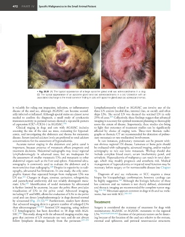
• Fig. 23.31 (A) The typical appearance of a large apocrine gland anal sac adenocarcinoma in a dog.
(B) The typical appearance of an apocrine gland anal sac adenocarcinoma in a cat. Ulceration with an
associated discharge is the most common finding in cats with apocrine gland anal sac adenocarcinomas.
is valuable for ruling out impaction, infection, or inflammatory Lymphadenopathy related to AGASAC can involve any of the
disease of the anal sac, although AGASAC can become second- three LN centers (medial iliac, internal iliac, or sacral), and often
arily infected or inflamed. Although special stains are almost never skips LNs. The sacral LN was deemed the sentinel LN in only
needed to confirm the diagnosis, a small study of cytokeratin 25% of cases. 635 Collectively, these findings suggest that advanced
immunoreactivity in perianal tumors showed a repeatable pattern imaging is necessary for optimal treatment planning to thoroughly
of expression (CK7+/CK14–) in AGASAC. 586 assess the extent of disease. Importantly, these studies also bring
Clinical staging in dogs and cats with AGASAC includes to light that outcomes of treatment studies can be significantly
assessing the size of the anal sac mass, evaluating for hypercal- affected by choice of staging tests. Three-view thoracic radio-
cemia, and investigating the abdomen and thorax for metastatic graphs or thoracic CT are recommended for detection of pulmo-
disease. Serum ionized calcium levels are preferred to total calcium nary metastasis or rare mediastinal involvement.
concentrations for the assessment of hypercalcemia. In rare instances, pulmonary metastasis can be present with-
Accurate tumor staging in the abdomen and pelvic canal is out obvious regional LN disease. Lameness or bone pain should
important, because presence of metastasis affects prognosis and be evaluated with radiography, advanced imaging, and/or nuclear
treatment decisions. Abdominal radiographs may reveal regional scintigraphy to rule out bone metastasis. Workup should also
lymphadenomegaly in advanced cases, but are inadequate for include complete blood count, serum biochemistry panel, and
the assessment of smaller metastatic LNs and metastasis to other urinalysis. Hypercalcemia of malignancy can result in renal dam-
abdominal organs such as the liver and spleen. Abdominal ultra- age, which may modify prognosis and anesthetic risk. Medical
sonography is commonly used to evaluate the abdomen and is management of hypercalcemia or impaired renal function may be
more sensitive than radiography. 630 Despite its superiority to radi- necessary before surgery or for nonresectable disease (see Chapter
ography, ultrasound has limitations. In one study, the only sono- 5).
graphic feature that separated benign from malignant LNs was Diagnosis of anal sac melanoma or SCC requires a tissue
LN size. 631 Changes in shape, contour, cavitation, echogenicity, biopsy for histopathologic confirmation; however, cytology can
and parenchymal uniformity did not reliably distinguish meta- be highly suggestive. 560 Although the biologic behavior of these
static LNs. 631 Identification of nodal metastasis by ultrasound less common anal sac tumors is not clearly defined, abdominal
is further limited by anatomy, because the pelvic floor precludes and thoracic imaging are recommended for complete tumor stag-
visualization of LNs in the pelvic canal. Advanced imaging, ing. 561–563 Metastasis appears common in dogs with anal sac mela-
including CT and MRI, allows for evaluation of LNs in the pelvic noma, but not with SCC. 560–563
canal and can detect lymphadenomegaly in dogs deemed normal
by ultrasound (Fig. 23.32). 632 Furthermore, studies have shown Treatment
that advanced imaging detects a greater number of enlarged LNs
than ultrasonograpy. 632–634 Sentinel LN mapping, using indirect Surgery is considered the mainstay of treatment for dogs with
CT lymphography, has been described in 18 dogs with AGA- nonmetastatic AGASAC or AGASAC metastatic to the regional
SAC. 635 This study, along with the advanced imaging studies, sug- LNs. 564,600,605,606,612 Excision of the primary tumor can be daunt-
gests that patterns of LN metastasis can vary, and do not always ing because of the location of the anal sacs relative to the rectum,
follow lymphatic drainage linearly from the perineum. 633–635 external anal sphincter, and perineal neurovascular structures;