
Page 1105 - Veterinary Immunology, 10th Edition
P. 1105
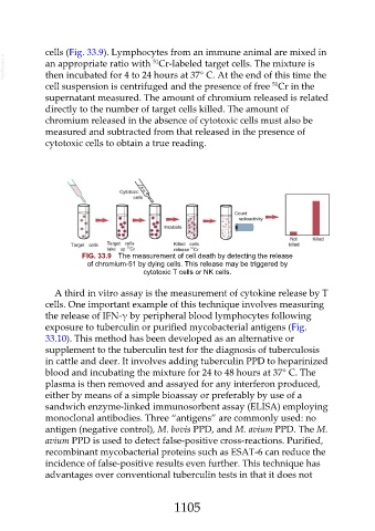
cells (Fig. 33.9). Lymphocytes from an immune animal are mixed in
  VetBooks.ir  an appropriate ratio with  Cr-labeled target cells. The mixture is
                                                  51
               then incubated for 4 to 24 hours at 37° C. At the end of this time the
                                                                                       51
               cell suspension is centrifuged and the presence of free  Cr in the
               supernatant measured. The amount of chromium released is related
               directly to the number of target cells killed. The amount of
               chromium released in the absence of cytotoxic cells must also be
               measured and subtracted from that released in the presence of

               cytotoxic cells to obtain a true reading.


                           FIG. 33.9  The measurement of cell death by detecting the release
                             of chromium-51 by dying cells. This release may be triggered by
                                               cytotoxic T cells or NK cells.


                  A third in vitro assay is the measurement of cytokine release by T

               cells. One important example of this technique involves measuring
               the release of IFN-γ by peripheral blood lymphocytes following
               exposure to tuberculin or purified mycobacterial antigens (Fig.
               33.10). This method has been developed as an alternative or
               supplement to the tuberculin test for the diagnosis of tuberculosis

               in cattle and deer. It involves adding tuberculin PPD to heparinized
               blood and incubating the mixture for 24 to 48 hours at 37° C. The
               plasma is then removed and assayed for any interferon produced,

               either by means of a simple bioassay or preferably by use of a
               sandwich enzyme-linked immunosorbent assay (ELISA) employing
               monoclonal antibodies. Three “antigens” are commonly used: no
               antigen (negative control), M. bovis PPD, and M. avium PPD. The M.
               avium PPD is used to detect false-positive cross-reactions. Purified,

               recombinant mycobacterial proteins such as ESAT-6 can reduce the
               incidence of false-positive results even further. This technique has
               advantages over conventional tuberculin tests in that it does not


                                                        1105