
Page 1106 - Veterinary Immunology, 10th Edition
P. 1106
sensitize the animal under testing by injection of antigen. In
VetBooks.ir addition, the animal does not have to be held for several days for
the test to be read. It is also much simpler than other in vitro tests
for cell-mediated immunity. The assay is at least as sensitive as the
single intradermal test and, if purified recombinant mycobacterial
proteins are employed, is highly specific. (Its sensitivity is about
85%, and its specificity is as high as 90% to 99%.) Positive results are
obtained earlier than by skin testing. However, it does appear to
detect tuberculosis in a slightly different population of animals than
the skin test. It has also been successfully used to diagnose Johne's
disease in sheep as well as tuberculosis in cats and dogs.
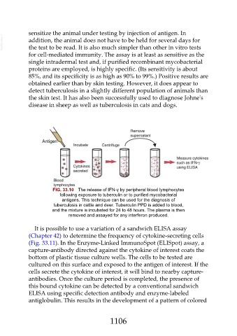
FIG. 33.10 The release of IFN-γ by peripheral blood lymphocytes
following exposure to tuberculin or to purified mycobacterial
antigens. This technique can be used for the diagnosis of
tuberculosis in cattle and deer. Tuberculin PPD is added to blood,
and the mixture is incubated for 24 to 48 hours. The plasma is then
removed and assayed for any interferon produced.
It is possible to use a variation of a sandwich ELISA assay
(Chapter 42) to determine the frequency of cytokine-secreting cells
(Fig. 33.11). In the Enzyme-Linked ImmunoSpot (ELISpot) assay, a
capture-antibody directed against the cytokine of interest coats the
bottom of plastic tissue culture wells. The cells to be tested are
cultured on this surface and exposed to the antigen of interest. If the
cells secrete the cytokine of interest, it will bind to nearby capture-
antibodies. Once the culture period is completed, the presence of
this bound cytokine can be detected by a conventional sandwich
ELISA using specific detection antibody and enzyme-labeled
antiglobulin. This results in the development of a pattern of colored
1106