
Page 147 - Veterinary Laser Therapy in Small Animal Practice
P. 147
Pointing light at musculoskeletal and neurological conditions: clinical applications 133
Case no. 13
B., canine, 3 months old, mixed breed, MNC, 7 kg
• Complaint: fracture.
• History: B. had been rescued after being hit by a car when he was 2 months old. He had a distal humeral
fracture, which had been repaired 3 days before, i.e. a month after the accident, through a medial approach,
using a clamp and rod internal fixation system and five screws. He was on cephalexin and carprofen.
• Physical examination: surgical wound over medial aspect of right humerus/elbow. Lameness score 4/5.
Severe tension in the muscles over the scapula and biceps. Carpus carried in flexion and withdrawal reflex
slightly delayed. VAS pain score 5/10.
• Diagnosis: postsurgical support for humeral fracture in a growing animal. Suspected radial neurapraxia.
• Treatment:
• Laser therapy:
• Treatment was started 3 days after surgery. Patient was treated twice a week initially and then once a
week, including scapula, humerus, elbow, and antebrachium, down to the carpus.
• A dose of 4–6 J/cm was used (only 2–4 initially over the wound) over a total of 200 cm ,with
2
2
passive ROM during treatment.
• Other:
• Cephalexin and carprofen maintained for 5 days.
• Outcome: carpal movement and posture was normal after two sessions and VAS dropped to 2/10 during the
second week. Two screws were removed in the fifth week after the initial surgery and the rest in the tenth
week. Notice the open growth plates in the proximal radius and anconeal process even 10 weeks after surgery
and after having been treated with LT (Fig. C13.4). At the moment of discharge, B. was able to run, even sprint
to the right side.
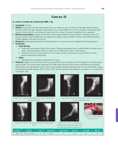
Figure C13.1 Fracture before surgery. (a) Craniocaudal and (b) Figure C13.2 Immediately after surgical repair. (a) Craniocaudal
mediolateral views. and (b) mediolateral views.
Figure C13.5 Patient during
treatment with LT.
Figure C13.3 Week 5. (a) Craniocaudal and (b) mediolateral Figure C13.4 Week 10, mediolateral
views. view. Note the open growth plates in the
proximal radius and anconeal process.
P (W) Tx time J/cm 2 Total J/Tx Spot (cm ) W/cm 2 Tx/week No. Tx
2
a
4 250 s (4.1 min) 5 1000 5 0.8 2-1 8
REDONDO PRINT (4-COL BLEED).indd 133 08/08/2019 09:48