
Page 157 - Veterinary Histology of Domestic Mammals and Birds, 5th Edition
P. 157
Blood and haemopoiesis (sanguis et haemocytopoesis) 139
The responding neutrophils phagocytose cell debris
VetBooks.ir and foreign material, perishing in the process. Lysosomal
enzymes are released into the interstitial tissue and cel-
lular components become incorporated into the process
of disintegration (pus formation). Thus, neutrophils (in
particular, among the granulocytes) are referred to as
microphages, as distinct from tissue macrophages.
Eosinophils (granulocytus eosinophilicus)
Eosinophils (Figure 7.6) are characterised by intensely aci-
dophilic (eosinophilic) granules. The size of the granules
varies with species (0.5–1.5 μm). They are particularly large
and prominent in the horse (Figure 7.13).
The granules are membrane-bound lysosomes con-
taining numerous enzymes, particularly catalases, acid
phosphatases, proteases, dehydrogenase and cathep-
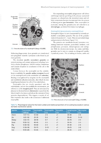
7.5 Fine structure of a neutrophil (dog; x10,000). sin. With the electron microscope, the canine and feline
granules can be seen to contain an elongated lamellar
Following phagocytosis, these granules are transformed crystalloid protein. This is lacking in horses, cattle and
into phagolytic vacuoles. Lysozyme is also found in pri-
mary granules.
The abundant specific (secondary) granules are
neutral-staining and contain substances including lacto-
ferrin, alkaline phosphatase, NADPH-oxidase, histaminase
and laminin receptors for attachment of the cell to the
endothelium.
Contact between the neutrophil and the endothe-
lium is established by specific surface receptors located
on the neutrophil and on the endothelial cell (selectins).
Binding is strengthened by integrins (on the neutrophil)
and endothelial adhesion molecules, after which the neu-
trophil passes between the cells of the endothelium.
Neutrophils that are closely associated with the
endothelium, and are thus available for extravasation, are
referred to as the marginal pool. They are stimulated by
physical or chemical factors (chemotaxis) to pass from the
blood vessel to the tissues and release the contents of their
granules (degranulation). This triggers a cascade of cel-
lular responses that leads to the attraction of additional
neutrophils. 7.6 Fine structure of an eosinophil (dog; x10,000).
Table 7.2 Physiological values for the total number and relative proportions of circulating leucocytes in various
domestic mammalian species.
Species Leucocytes Neutrophils Eosinophils Basophils Lymphocytes Monocytes
1000/μl (%) (%) (%) (%) (%)
Horse 9 (7–11) 52–60 2–4 <1 30–40 3–4
Ox 8 (5–10) 25–35 5–6 <1 55–65 5–10
Sheep 8 (6–12) 30–40 5–7 <1 45–70 2–5
Goat 10 (8–12) 40–45 3–5 <1 50–55 3–5
Pig 12 (8–16) 50–60 2–3 <1 35–50 2–6
Dog 12 (8–18) 55–75 3–10 <1 20–25 2–6
Cat 10 (9–24) 55–65 3–6 <1 30–35 2–5
Vet Histology.indb 139 16/07/2019 14:58